

Axial Loading during MRI Induces Lumbar Foraminal Area Changes and Has the Potential to Improve Diagnostics of Nerve Root Compromise
- PMID: 35456215
- PMCID: PMC9029659
- DOI: 10.3390/jcm11082122
Axial Loading during MRI Induces Lumbar Foraminal Area Changes and Has the Potential to Improve Diagnostics of Nerve Root Compromise
Abstract
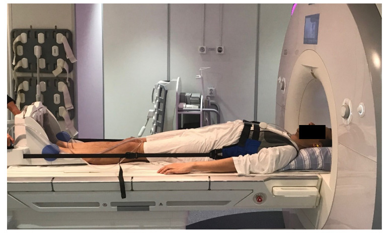
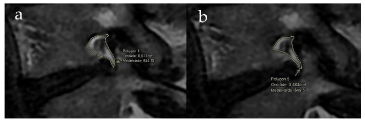
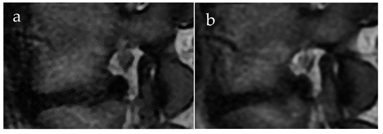
Lumbar foraminal stenosis is a common cause of lumbar radiculopathy and conventionally assessed with magnetic resonance imaging (MRI) in supine-positioned patients. An MRI acquired during spine loading may unmask pathology not otherwise revealed in a relaxed position. Therefore, we investigated how spine loading during MRI affects lumbar foramina. In 89 low-back pain patients' lumbar, MRIs were performed in a relaxed supine position and during axial loading using a Dynawell® compression device. The smallest area of all intervertebral foramina at levels L3/L4-L5/S1 (534 foramina) was determined using a freehand polygonal tool in parasagittal T2-weighted sequences. The grading system described by Lee et al. was also used to qualitatively assess foraminal stenosis. Overall, a mean reduction of 2.2% (mean -0.89 cm2 and -0.87 cm2, respectively) was observed (p = 0.002), however for individual foramina large variations, with up to about 50% increase or decrease, were seen. Stratified for lumbar level, an area reduction was found for L3/L4 and L4/L5 foramina (mean change -0.03 cm2; p = 0.036; and -0.03 cm2; p = 0.004, respectively) but not for L5/S1. When comparing the measured area changes to qualitative foraminal grading, 22% of the foramina with a measured area decrease were evaluated with a higher grading. Thus, detailed information on foraminal appearance and nerve root affection can be obtained using this method.
Keywords: axial loading during MRI; diagnostics; lumbar foraminal area; lumbar foraminal stenosis.
Conflict of interest statement
The authors declare no conflict of interest.
Figures
References
-
- Splendiani A., Perri M., Grattacaso G., Di Tunno V., Marsecano C., Panebianco L., Gennarelli A., Felli V., Varrassi M., Barile A., et al. Magnetic resonance imaging (MRI) of the lumbar spine with dedicated G-scan machine in the upright position: A retrospective study and our experience in 10 years with 4305 patients. Radiol. Med. 2016;121:38–44. doi: 10.1007/s11547-015-0570-9. - DOI - PubMed
Grants and funding
LinkOut - more resources
Full Text Sources
Medical
