. 2022 Apr 12;11(8):2156.
doi: 10.3390/jcm11082156.

Rapid Campimetry-A Novel Screening Method for Glaucoma Diagnosis

Affiliations

Rapid Campimetry-A Novel Screening Method for Glaucoma Diagnosis

Fabian Müller et al. J Clin Med. .

Abstract

One of the most important functions of the retina-the enabling of perception of fast movements-is largely suppressed in standard automated perimetry (SAP) and kinetic perimetry (Goldmann) due to slow motion and low contrast between test points and environment. Rapid campimetry integrates fast motion (=10°/4.7 s at 40 cm patient-monitor distance) and high contrast into the visual field (VF) examination in order to facilitate the detection of absolute scotomas. A bright test point moves on a dark background through the central 10° VF. Depending on the distance to the fixation point, the test point automatically changes diameter (≈0.16° to ≈0.39°). This method was compared to SAP (10-2 program) for six subjects with glaucoma. Rapid campimetry proved to be comparable and possibly better than 10-2 SAP in identifying macular arcuate scotomas. In four subjects, rapid campimetry detected a narrow arcuate absolute scotoma corresponding to the nerve fiber course, which was not identified as such with SAP. Rapid campimetry promises a fast screening method for the detection of absolute scotomas in the central 10° visual field, with a potential for cloud technologies and telemedical applications. Our proof-of-concept study motivates systematic testing of this novel method in a larger cohort.

Keywords: arcuate scotoma; automated static perimetry; glaucoma; rapid campimetry; telemedicine; visual field defect.


Conflict of interest statement

Fabian Müller received no support for the present manuscript but he had funding for the patenting with H & M Medical Solutions GmbH. Other authors had none. The authors declare no conflict of interest.

Figures
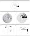
Figure 1
Figure 1
F.H. Visual field findings. (a) Sketch of the grey, drawn scotoma in the visual field of the right eye subjectively perceived by F.H. (first scotoma—one arrow, second scotoma—two arrows). (b) 30° field of view examined with Octopus 301. The scotoma is dark grey in the temporal upper field of view, the blind spot is shown in white. (c) 10° central visual field examined with the Humphrey Field Analyzer (HFA3). The absolute scotoma is black in the temporal superior visual field, and the relative scotoma is dark grey in the temporal and nasal superior visual fields. (d) Red and green dots, connected by a grey line, represent the beginning and end of the scotoma in the paramacular visual field after the screening procedure. One arrow marks the first scotoma, two arrows marks the second. (e) The 15° central visual field findings of scotoma delineation campimetry. After finding the two scotomas in the screening procedure, the exact scotoma borders were determined. The four grey spots between the arcuate scotoma and blind spot (black) represent the presumed scotoma course. In this area, the test point is thicker than the narrow scotoma and therefore does not become invisible. When the test dot moves quickly, a brightness difference is perceived here, indicating the defect.
Figure 2
Figure 2
Rapid campimetry testing environment. Left Panel: Snapshot of the actual campimetry setting (with increased room lighting for better visualisation) with a volunteer fixating the centre of the testing area; left part of the image is masked to disable identification. Right panel: a sketch showing a person (P) looking at the monitor with a 40 cm distance (A) while an examiner (E) controls and runs the test on a different monitor.
Figure 3
Figure 3
The path tested in the screening procedure of rapid campimetry is shown in dashed lines. In the arc scotoma marked by a black ring, 8° from the fixation point, the optimum test point speed was determined. The test point changes its diameter with the distance from the fixation point; the greater the distance, the greater its diameter. In the outer of the three vertical test lines, the used size change is overdrawn. If one wants to determine the examined area in this area, then the area is calculated as the sum of two identical trapezoids.
Figure 4
Figure 4
Eyes with normal visual field of subjects 1–5. The blind spot was detectable at 15° for all subjects except subject 2 (S2) with rapid campimetry. SAP = standard automated perimetry. OD = right eye; OS = left eye; S: subject; SAP: standard automated perimetry.
Figure 5
Figure 5
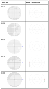
Eyes with visual field defects of subjects 1–5 compared between 10-2 SAP vs. rapid campimetry and scotoma delineation campimetry. SAP = standard automated perimetry. OD = right eye; OS = left eye; S: subject.

References

    1. Tham Y.-C., Li X., Wong T.Y., Quigley H.A., Aung T., Cheng C.-Y. Global Prevalence of Glaucoma and Projections of Glaucoma Burden through 2040: A Systematic Review and Meta-Analysis. Ophthalmology. 2014;121:2081–2090. doi: 10.1016/j.ophtha.2014.05.013. - DOI - PubMed
    1. Al-Nosairy K.O., Hoffmann M.B., Bach M. Non-invasive Electrophysiology in Glaucoma, Structure and Function—A Review. Eye. 2021;35:2374–2385. doi: 10.1038/s41433-021-01603-0. - DOI - PMC - PubMed
    1. Chauhan B.C., Garway-Heath D.F., Goñi F.J., Rossetti L., Bengtsson B., Viswanathan A.C., Heijl A. Practical Recommendations for Measuring Rates of Visual Field Change in Glaucoma. Br. J. Ophthalmol. 2008;92:569–573. doi: 10.1136/bjo.2007.135012. - DOI - PMC - PubMed
    1. Nakanishi M., Wang Y.-T., Jung T.-P., Zao J.K., Chien Y.-Y., Diniz-Filho A., Daga F.B., Lin Y.-P., Wang Y., Medeiros F.A. Detecting Glaucoma with a Portable Brain-Computer Interface for Objective Assessment of Visual Function Loss. JAMA Ophthalmol. 2017;135:550–557. doi: 10.1001/jamaophthalmol.2017.0738. - DOI - PMC - PubMed
    1. Wu Z., Medeiros F.A. Recent Developments in Visual Field Testing for Glaucoma. Curr. Opin. Ophthalmol. 2018;29:141–146. doi: 10.1097/ICU.0000000000000461. - DOI - PubMed

LinkOut - more resources