Case Reports
. 2022 Apr 9;13(4):662.
doi: 10.3390/genes13040662.

Phenotype Expansion for Atypical Gaucher Disease Due to Homozygous Missense PSAP Variant in a Large Consanguineous Pakistani Family

Affiliations
Case Reports

Phenotype Expansion for Atypical Gaucher Disease Due to Homozygous Missense PSAP Variant in a Large Consanguineous Pakistani Family

Khurram Liaqat et al. Genes (Basel). .

Abstract

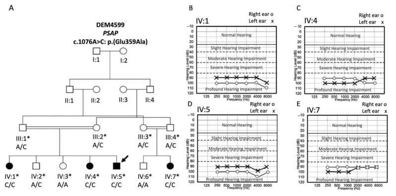
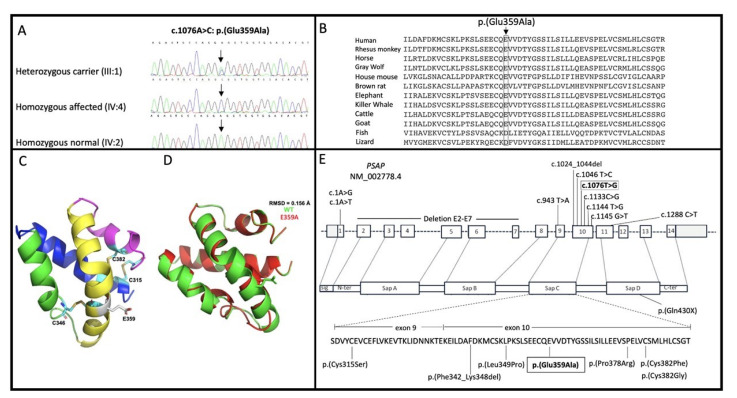
Atypical Gaucher disease is caused by variants in the PSAP gene. Saposin C is one of four homologous proteins derived from sequential cleavage of the saposin precursor protein, prosaposin. It is an essential activator for glucocerebrosidase, which is deficient in Gaucher disease. Although atypical Gaucher disease due to deficiency of saposin C is rare, it exhibits vast phenotypic heterogeneity. Here, we report on a Pakistani family that exhibits features of Gaucher disease, i.e., prelingual profound sensorineural hearing impairment, vestibular dysfunction, hepatosplenomegaly, kyphosis, and thrombocytopenia. The family was investigated using exome and Sanger sequencing. A homozygous missense variant c.1076A>C: p.(Glu359Ala) in exon 10 of the PSAP gene was observed in all affected family members. In conclusion, we identified a new likely pathogenic missense variant in PSAP in a large consanguineous Pakistani family with atypical Gaucher disease. Gaucher disease due to a deficiency of saposin C has not been previously reported within the Pakistani population. Genetic screening of patients with the aforementioned phenotypes could ensure adequate follow-up and the prevention of further complications. Our finding expands the genetic and phenotypic spectrum of atypical Gaucher disease due to a saposin C deficiency.

Keywords: atypical Gaucher disease; exome sequencing; hearing impairment; saposin C.


Conflict of interest statement

The authors declare no conflict of interest.

Figures

Figure 1
Figure 1
Pedigree and audiograms for family DEM4599. (A) Pedigree drawing of family DEM4599 segregating GD with an autosomal recessive mode of inheritance. Circles represent females and squares males. Filled symbols indicate affected family members and clear symbols unaffected members. Double lines in the pedigree represent a consanguineous marriage. Asterisks indicate individuals for whom a DNA sample is available, and an arrow indicates the sample selected for exome sequencing. Below each family member the PSAP c.1076A>C genotype is displayed. Audiograms of the affected individuals of the family DEM4599 (B) IV:1 (12 years of age), (C) IV:4 (14 years of age), (D) IV:5 (20 years of age), and (E) IV:7 (19 years of age).
Figure 2
Figure 2
Overview of PSAP (NM_002778.4) protein domains, sequence data on variant c.1076A>C, p.(Glu359Ala) in family DEM4599 and a predicted three-dimensional structure of Sap-C. (A) Chromatograms showing likely pathogenic missense variant c.1076A>C, p.(Glu359Ala) in the PSAP in heterozygous unaffected carrier (III:1) (upper panel), homozygous affected family member (IV:4) (middle panel), and homozygous wild-type unaffected family member individual (IV:2) (bottom panel). (B) The mutated amino acid residue Glu359 located in the Sap-C domain is conserved among various species analyzed. (C) Three-dimensional structure of the wild-type Sap-C domain in ribbon representation with the conserved cysteine residues are indicated. (D) The superposed structure of wild (green color) and mutant (red color) structure showing the difference in overall confirmation due to the p.E359A mutation. RMSD (root mean square deviation). (E) Structure of PSAP (NM_002778.4) and the protein it encodes. The protein is composed of Sap A-D domains. The missense variant c.1076A>C, p.(Glu359Ala) located in exon 10 of the PSAP (NM_002778.4) identified in this study is displayed in a box. All other variants which have been identified for atypical GD are indicated by a line. Exons 9 and 10 of the PSAP encode the Sap-C domain. Besides c.1076A>C, p.(Glu359Ala) the only other variant that was previously observed in the Sap-C domain in the homozygous state is p.(Phe342_Lys348del).

Similar articles

Cited by

References

    1. Tajima A., Yokoi T., Ariga M., Ito T., Kaneshiro E., Eto Y., Ida H. Clinical and genetic study of Japanese patients with type 3 Gaucher disease. Mol. Genet. Metab. 2009;97:272–277. doi: 10.1016/j.ymgme.2009.05.001. - DOI - PubMed
    1. Mistry P., Smith S., Ali M., Cox T., Hatton C., McIntyre N. Genetic diagnosis of Gaucher’s disease. Lancet. 1992;339:889–892. doi: 10.1016/0140-6736(92)90928-V. - DOI - PubMed
    1. Graves P.N., Grabowski G.A., Eisner R., Palese P., Smith F.I. Gaucher Disease Type 1: Cloning and Characterization of a cDNA Encoding Acid β-Glucosidase from an Ashkenazi Jewish Patient. DNA. 1988;7:521–528. doi: 10.1089/dna.1.1988.7.521. - DOI - PubMed
    1. Chérin P., Rose C., De Roux-Serratrice C., Tardy D., Dobbelaere D., Grosbois B., Hachulla E., Jaussaud R., Javier R.-M., Noël E., et al. The neurological manifestations of Gaucher disease type 1: The French Observatoire on Gaucher disease (FROG) J. Inherit. Metab. Dis. 2010;33:331–338. doi: 10.1007/s10545-010-9095-5. - DOI - PubMed
    1. Tylki-Szymańska A., Czartoryska B., Vanier M.-T., Poorthuis B., Groener J., Ługowska A., Millat G., Vaccaro A., Jurkiewicz E. Non-neuronopathic Gaucher disease due to saposin C deficiency. Clin. Genet. 2007;72:538–542. doi: 10.1111/j.1399-0004.2007.00899.x. - DOI - PubMed

Publication types