

Niosomal Nanocarriers for Enhanced Dermal Delivery of Epigallocatechin Gallate for Protection against Oxidative Stress of the Skin
- PMID: 35456560
- PMCID: PMC9029719
- DOI: 10.3390/pharmaceutics14040726
Niosomal Nanocarriers for Enhanced Dermal Delivery of Epigallocatechin Gallate for Protection against Oxidative Stress of the Skin
Abstract
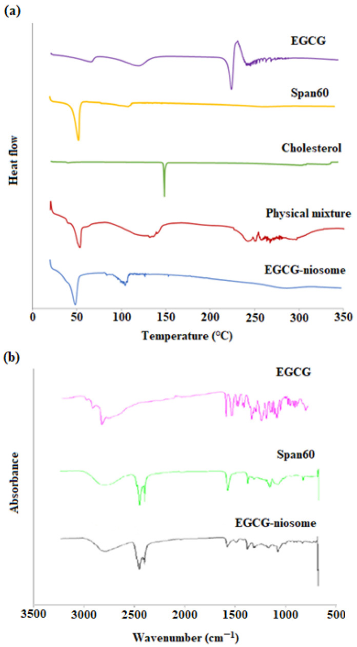
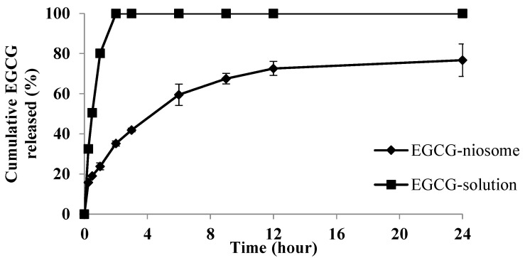
Among green tea catechins, epigallocatechin gallate (EGCG) is the most abundant and has the highest biological activities. This study aims to develop and statistically optimise an EGCG-loaded niosomal system to overcome the cutaneous barriers and provide an antioxidant effect. EGCG-niosomes were prepared by thin film hydration method and statistically optimised. The niosomes were characterised for size, zeta potential, morphology and entrapment efficiency. Ex vivo permeation and deposition studies were conducted using full-thickness human skin. Cell viability, lipid peroxidation, antioxidant enzyme activities after UVA-irradiation and cellular uptake were determined. The optimised niosomes were spherical and had a relatively uniform size of 235.4 ± 15.64 nm, with a zeta potential of -45.2 ± 0.03 mV and an EE of 53.05 ± 4.46%. The niosomes effectively prolonged drug release and demonstrated much greater skin penetration and deposition than free EGCG. They also increased cell survival after UVA-irradiation, reduced lipid peroxidation, and increased the antioxidant enzymes' activities in human dermal fibroblasts (Fbs) compared to free EGCG. Finally, the uptake of niosomes was via energy-dependent endocytosis. The optimised niosomes have the potential to be used as a dermal carrier for antioxidants and other therapeutic compounds in the pharmaceutical and cosmetic industries.
Keywords: antioxidant activity; catechin; cellular uptake; dermal delivery; niosomes; oxidative stress; penetration; skin barrier.
Conflict of interest statement
The authors declare no conflict of interest.
Figures


References
-
- Hemrajani C., Negi P., Parashar A., Gupta G., Jha N.K., Singh S.K., Chellappan D.K., Dua K. Overcoming drug delivery barriers and challenges in topical therapy of atopic dermatitis: A nanotechnological perspective. Biomed. Pharmacother. 2022;147:112633. doi: 10.1016/j.biopha.2022.112633. - DOI - PubMed
-
- Ng K.W., Lau W.M. Percutaneous Penetration Enhancers Chemical Methods in Penetration Enhancement. Springer; Berlin, Germany: 2015. Skin Deep: The Basics of Human Skin Structure and Drug Penetration; pp. 1–7.
LinkOut - more resources
Full Text Sources
