. 2022 Apr 6;14(4):802.
doi: 10.3390/pharmaceutics14040802.

Regulation of p27 (Kip1) by Ubiquitin E3 Ligase RNF6

Affiliations

Regulation of p27 (Kip1) by Ubiquitin E3 Ligase RNF6

Dhanraj Deshmukh et al. Pharmaceutics. .

Abstract

The cyclin-dependent kinase inhibitor p27 (Kip1) is an important regulator of the G1/S checkpoint. It is degraded by the SCF-SKP2 complex in late G1 thereby allowing cells to progress to the S phase. Here we investigated the role of the E3 ubiquitin ligase RNF6 (Ring Finger Protein 6) in cell cycle progression in prostate cancer cells. Our data demonstrate that RNF6 can promote cell cycle progression by reducing the levels of p27. Knockdown of RNF6 led to an increase in the stability of p27 and to the arrest of cells in the G1 phase. RNF6 interacted with p27 via its KIL domain and this interaction was found to be phosphorylation independent. RNF6 enhanced ubiquitination and subsequent degradation of p27 in the early G0/G1 phase of the cell cycle. Knockdown of RNF6 expression by short hairpin RNA led to inhibition of the CDK2/Cyclin E complex thereby reducing phosphorylation of Retinoblastoma protein (Rb) and to a subsequent decrease in cell cycle progression and proliferation. Our data suggest that RNF6 acts as a negative regulator for p27kip1 leading to its proteasome-dependent degradation in the early G0/G1 phase of the cell cycle.

Keywords: RNF6; cell cycle; p27; prostate cancer.


Conflict of interest statement

The author (Jin Xu) is currently an employee of AstraZeneca, however, he did not work for AstraZeneca at the time of experiments and manuscript preparation. The other authors declare no conflict of interest.

Figures

Figure 1
Figure 1
Knockdown of RNF6 leads to increase in stability of p27. (A) Prostate cancer cell lines were transduced with either control shRNA or shRNF6-1/2. After 72 h of knockdown, cells were lysed and lysates subjected to western blot analyses. (B) LNCaP cells were transduced with either control shRNA or shRNF6 1/2. After 72 h, cells were lysed and lysates subjected to Western blot analysis. (C) HEK293 cells were transfected with the indicated plasmids by the CaCl2 method. After 42 h, MG132 (10 µM) was added as shown. After 48 h of transfection, cells were lysed in RIPA buffer with protease inhibitors. The lysates were then subjected to western blot analysis.
Figure 2
Figure 2
RNF6 interacts with p27 in a phosphorylation independent manner. (A) Prostate cancer cells were treated with MG132 (10 µM) for 6 h and then lysed in IP buffer. Lysates were immunoprecipitated with anti-RNF6 antibody and immunoblotted for p27. Preimmune IgG was used as control. (B) LNCaP and CWR-R1 cells were transduced with either control or RNF6 shRNA. After about 66 h post-transduction, cells were treated with MG132 (10 µM) for 6 h. Cells were then lysed in IP buffer and the lysates immunoprecipitated with anti-RNF6 antibody. Immunoprecipitates were then immunoblotted for p27. RNF6 blots were used as control. (C) Immunofluorescence confocal microscopy was carried out in CWR-R1 cells with anti-RNF6 (Red) and anti-p27 (green) antibodies. Nuclei were counterstained with DAPI. (D) LNCaP cells were transfected with either GST or GST-p27 alone or with RNF6. After 48 h of transfection, cell lysates were prepared and GST immunoprecipitated with GSH beads. The immunoprecipitate was then immunoblotted for GST and RNF6. (E) HA-tagged WT RNF6 and various mutants were cotransfected with p27 in HEK293 cells. After 42 h, cells were treated with MG132 (10 µM) for 6 h after which they were lysed and lysates immunoblotted for HA and p27. Cartoons of WT RNF6 and different mutants. (F) WT RNF6 or Myc-tagged SKP2 was cotransfected with WT p27 or the phosphomutant p27T187A in HEK293 cells. Cells were treated with MG132 (10 µM) for 6 h prior to cell lysis. After 48 h of transfection, cells were lysed and lysates immunoprecipitated with anti-RNF6 antibody. Immunoprecipitates were subjected to western blot analysis and immunoblotted for HA/Myc and p27.
Figure 3
Figure 3
RNF6 knockdown leads to increase in p27 levels in the nucleus. (A) CWR-R1 cells were transduced with either control shRNA or shRNF6. After 72 h, cells were fixed, and immunofluorescence confocal microscopy was carried out on the cells by co-staining with anti-p27 (green) and anti-RNF6 (red) antibodies. Cells were counterstained with DAPI. (B) LNCaP/CWR-R1 cells were transduced with either control shRNA or shRNF6. After about 72 h of transduction, cells were fractionated as described in the Materials and Methods. The cytosolic and nuclear fractions were then subjected to Western blot analysis.
Figure 4
Figure 4
RNF6 ubiquitinates p27 in vivo and in vitro. (A) LNCaP cells were transduced with scrambled shRNA, shRNF6, or RNF6 plasmid. After about 72 h of transduction, cells were lysed and lysates immunoprecipitated with anti-p27 antibody. Immunoprecipitates were then probed for Ub. (B) HEK293 cells were transfected with either p27 alone, WT-RNF6 and p27, or mutant RNF6 (ΔRING) and p27. After 42 h of transfection, cells were treated with MG132 for an additional 6 h. Cells were then lysed in denaturing buffer and lysates immunoprecipitated with anti-p27 antibody. The immunoprecipitates were immunoblotted for Ub. (C) The in vitro ubiquitination assays were carried out as described in Materials and Methods. Top, immunoblot of anti-p27 to detect p27. Bottom: Coomassie blue staining (CBS) of the gel to monitor the amount of E3 ligases present in the reactions. (D) Same as in C except that either active or inactive GST-RNF6 was used.
Figure 5
Figure 5
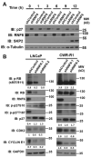
RNF6 knockdown leads to p27 accumulation in early G1 and reduced Rb phosphorylation. (A) CWR-R1 cells were transduced wither either control sRNA or shRNF6. After 48 h of transduction, cells were synchronized in G0/G1 by serum withdrawal for 48 h. Ten percent final serum was added back and cells were harvested at indicated time points. Cells lysates were prepared and immunoblotted as shown. (B) CWR-R1 and LNCaP cells were transduced with either control shRNA or shRNF6. After 72 h of transduction, cell lysates were prepared and subjected to Western blot analysis as shown.
Figure 6
Figure 6
Knockdown of RNF6 leads to G1/S block and to reduced cell proliferation. (A) Asynchronous cells were transduced with either control shRNA or shRNF6 for 72 h after which they were stained with PI as described in materials and methods. After PI staining, cells were subjected to FACS analysis. (B) Cells were transduced with either control shRNA or RNF6 shRNA for 7 days. Cell proliferation was then assessed using CCK8 assay, * p < 0.05. (C) Cells were transduced with control or RNF6 shRNA as in B. Cells were then fixed and stained with Coomassie blue dye.

References

    1. Jiao B., Taniguchi-Ishigaki N., Güngör C., Peters M.A., Chen Y.-W., Riethdorf S., Drung A., Ahronian L.G., Shin J., Pagnis R., et al. Functional activity of RLIM/Rnf12 is regulated by phosphorylation-dependent nucleocytoplasmic shuttling. Mol. Biol. Cell. 2013;24:3085–3096. doi: 10.1091/mbc.e13-05-0239. - DOI - PMC - PubMed
    1. Macdonald D.H., Lahiri D., Sampath A., Chase A., Sohal J., Cross N.C. Cloning and characterization of RNF6, a novel RING finger gene mapping to 13q12. Genomics. 1999;58:94–97. doi: 10.1006/geno.1999.5802. - DOI - PubMed
    1. Tursun B., Schlüter A., Peters M.A., Viehweger B., Ostendorff H.P., Soosairajah J., Drung A., Bossenz M., Johnsen S.A., Schweizer M., et al. The ubiquitin ligase Rnf6 regulates local LIM kinase 1 levels in axonal growth cones. Genes Dev. 2005;19:2307–2319. doi: 10.1101/gad.1340605. - DOI - PMC - PubMed
    1. Xu K., Shimelis H., Linn D.E., Jiang R., Yang X., Sun F., Guo Z., Chen H., Li W., Chen H., et al. Regulation of Androgen Receptor Transcriptional Activity and Specificity by RNF6-Induced Ubiquitination. Cancer Cell. 2009;15:270–282. doi: 10.1016/j.ccr.2009.02.021. - DOI - PMC - PubMed
    1. Takahashi H., Uematsu A., Yamanaka S., Imamura M., Nakajima T., Doi K., Yasuoka S., Takahashi C., Takeda H., Sawasaki T. Establishment of a Wheat Cell-Free Synthesized Protein Array Containing 250 Human and Mouse E3 Ubiquitin Ligases to Identify Novel Interaction between E3 Ligases and Substrate Proteins. PLoS ONE. 2016;11:e0156718. doi: 10.1371/journal.pone.0156718. - DOI - PMC - PubMed

LinkOut - more resources