. 2022 Mar 25;10(4):710.
doi: 10.3390/microorganisms10040710.

Absence of Streptococcus pneumoniae Capsule Increases Bacterial Binding, Persistence, and Inflammation in Corneal Infection

Affiliations

Absence of Streptococcus pneumoniae Capsule Increases Bacterial Binding, Persistence, and Inflammation in Corneal Infection

Mary A Carr et al. Microorganisms. .

Abstract

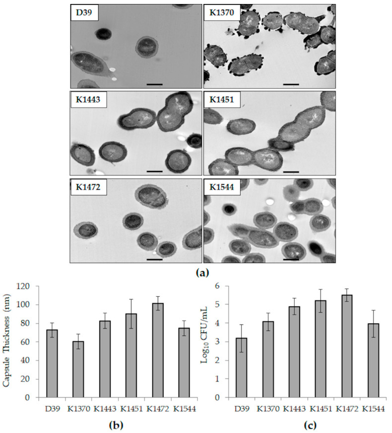
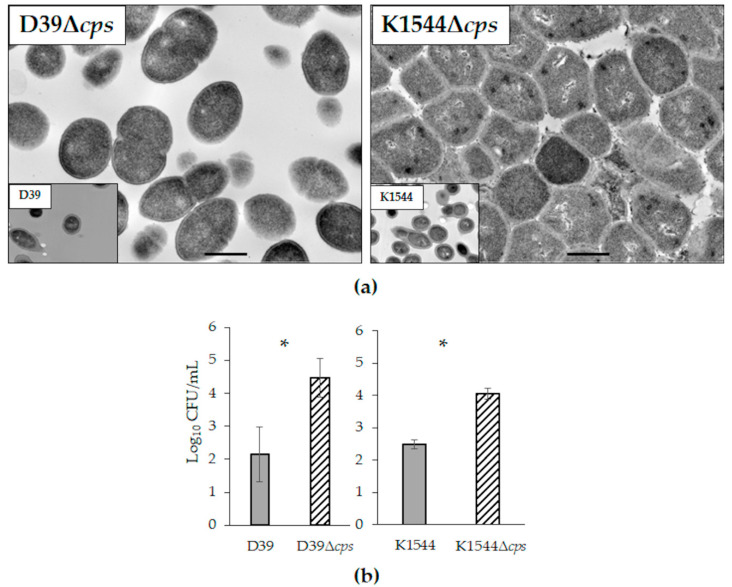
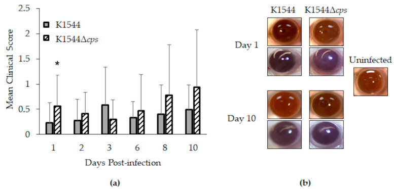
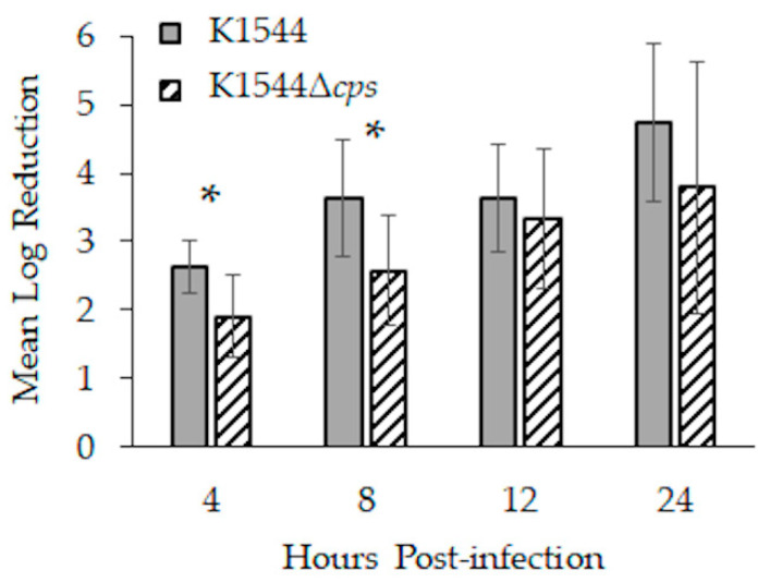
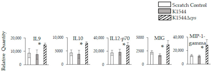
The role of the pneumococcal polysaccharide capsule is largely unclear for Streptococcus pneumoniae keratitis, an ocular inflammatory disease that develops as a result of bacterial infection of the cornea. In this study, capsule-deficient strains were compared to isogenic parent strains in their ability to adhere to human corneal epithelial cells. One isogenic pair was further used in topical ocular infection of mice to assess the contribution of the capsule to keratitis. The results showed that non-encapsulated pneumococci were significantly more adherent to cells, persisted in significantly higher numbers on mouse corneas in vivo, and caused significant increases in murine ocular IL9, IL10, IL12-p70, MIG, and MIP-1-gamma compared to encapsulated S. pneumoniae. These findings indicate that the bacterial capsule impedes virulence and the absence of capsule impacts inflammation following corneal infection.

Keywords: Streptococcus pneumoniae; adhesion; capsule; inflammation; ocular; pneumococcus.


Conflict of interest statement

The authors declare no conflict of interest.

Figures

Figure 1
Figure 1
Capsules thickness and relative host cell adhesion of the strains included in this study. (a) TEM images of each strain. The bar in each panel represents a length of 500 nm; (b) average capsule thicknesses as measured from TEM images; (c) average log CFU/mL bound to HCECs. Error bars represent standard deviation.
Figure 2
Figure 2
Isogenic non-encapsulated mutants of strains D39 and K1544. (a) TEM images of each non-encapsulated strain, with an inset of the parent strain for comparison. The bar in each panel represents a length of 500 nm; (b) average log CFU/mL bound to HCECs. Error bars represent standard deviation. Each asterisk indicates a significant difference between parent and mutant.
Figure 3
Figure 3
Keratitis in mice. (a) Mean clinical scores as determined by degree of corneal opacity on a scale of 0 to 4. Error bars represent standard deviation. Asterisk indicates a significant difference between parent and mutant; (b) representative photographs (2 for each strain at each time point shown). Corneal opacity was nearly undetectable for eyes infected with K1544, as demonstrated by clear visualization of pupils. For those infected with K1544Δcps, opacity was detected as shown by slight obscurity of the pupils (bottom eye for Day 1 and bottom eye for Day 10).
Figure 4
Figure 4
Bacterial load reductions during the first 24 h of infection in mice. Corneas were infected with approximately 8 log10 CFU, and each data bar indicates by how many log10 CFU that starting inoculum decreased. Error bars represent standard deviation. Asterisks indicate a significant difference between parent and mutant.
Figure 5
Figure 5
Inflammation-associated markers in mouse eyes 24 h after infection. Relative quantities are expressed as intensity values generated by ImageJ software. Error bars represent standard deviation. Asterisks indicate a significant difference between parent and mutant. Significant differences were detected only at 24 h.

References

    1. Shayegani M., Parsons L.M., Gibbons W.E., Jr., Campbell D. Characterization of nontypable Streptococcus pneumoniae-like organisms isolated from outbreaks of conjunctivitis. J. Clin. Microbiol. 1982;16:8–14. doi: 10.1128/jcm.16.1.8-14.1982. - DOI - PMC - PubMed
    1. Carvalho M.G., Steigerwalt A.G., Thompson T., Jackson D., Facklam R.R. Confirmation of nontypeable Streptococcus pneumoniae-like organisms isolated from outbreaks of epidemic conjunctivitis as Streptococcus pneumoniae. J. Clin. Microbiol. 2003;41:4415–4417. doi: 10.1128/JCM.41.9.4415-4417.2003. - DOI - PMC - PubMed
    1. Haas W., Hesje C.K., Sanfilippo C.M., Morris T.W. High proportion of nontypeable Streptococcus pneumoniae isolates among sporadic, nonoutbreak cases of bacterial conjunctivitis. Curr. Eye Res. 2011;36:1078–1085. doi: 10.3109/02713683.2011.624670. - DOI - PubMed
    1. Martin M., Turco J.H., Zegans M.E., Facklam R.R., Sodha S., Elliott J.A., Pryor J.H., Beall B., Erdman D.D., Baumgartner Y.Y., et al. An outbreak of conjunctivitis due to atypical Streptococcus pneumoniae. N. Engl. J. Med. 2003;348:1112–1121. doi: 10.1056/NEJMoa022521. - DOI - PubMed
    1. Porat N., Greenberg D., Givon-Lavi N., Shuval D.S., Trefler R., Segev O., Hanage W.P., Dagan R. The important role of nontypable Streptococcus pneumoniae international clones in acute conjunctivitis. J. Infect. Dis. 2006;194:689–696. doi: 10.1086/506453. - DOI - PubMed