. 2022 Apr 12;23(8):4262.
doi: 10.3390/ijms23084262.

A Novel NOX Inhibitor Treatment Attenuates Parkinson's Disease-Related Pathology in Mouse Models

Affiliations

A Novel NOX Inhibitor Treatment Attenuates Parkinson's Disease-Related Pathology in Mouse Models

Anurupa A Ghosh et al. Int J Mol Sci. .

Abstract

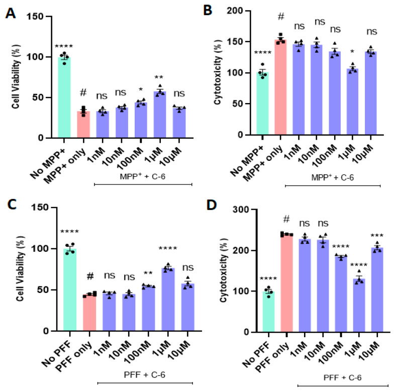
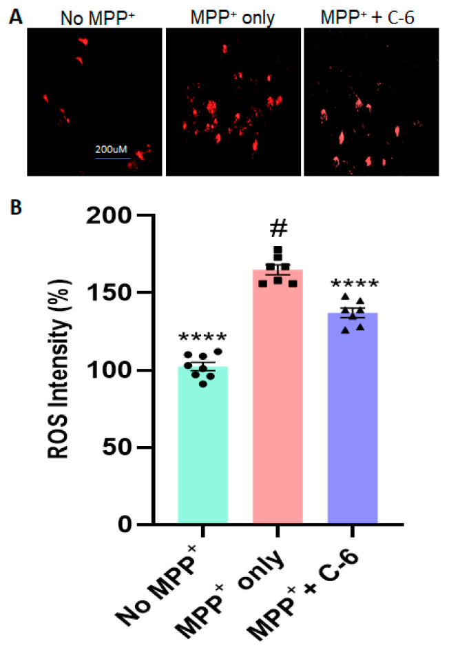
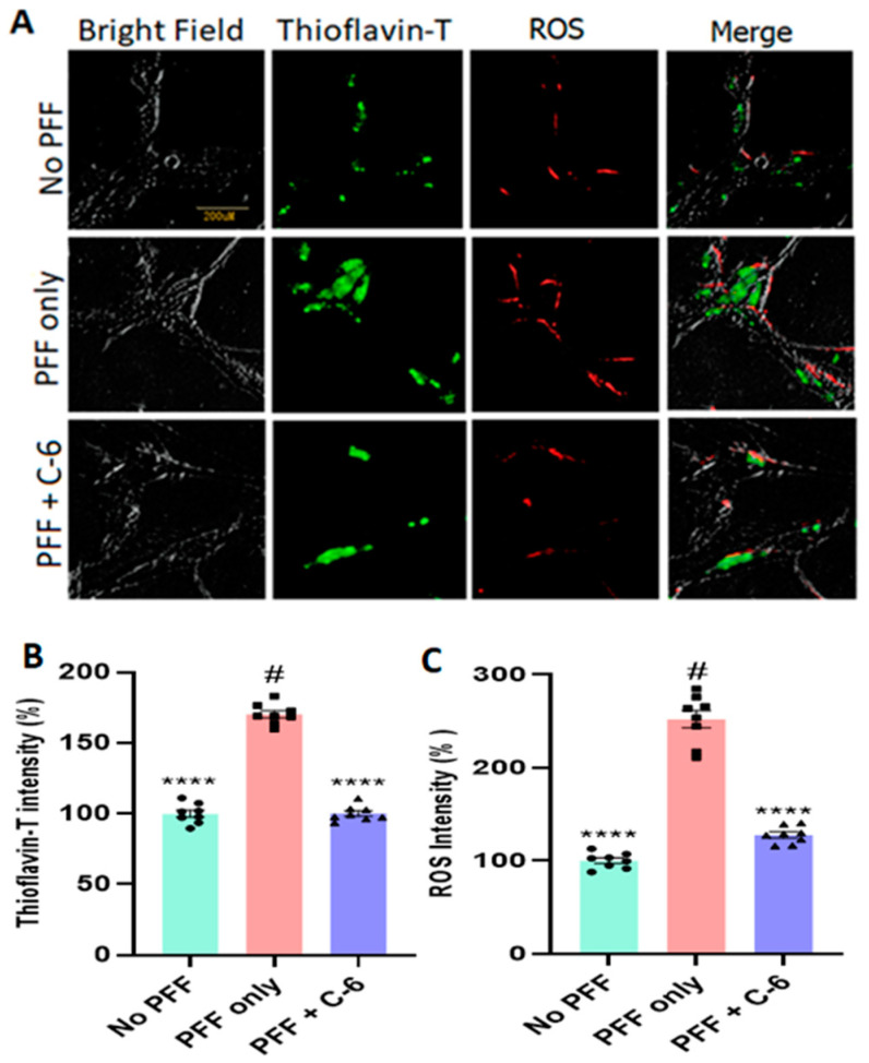
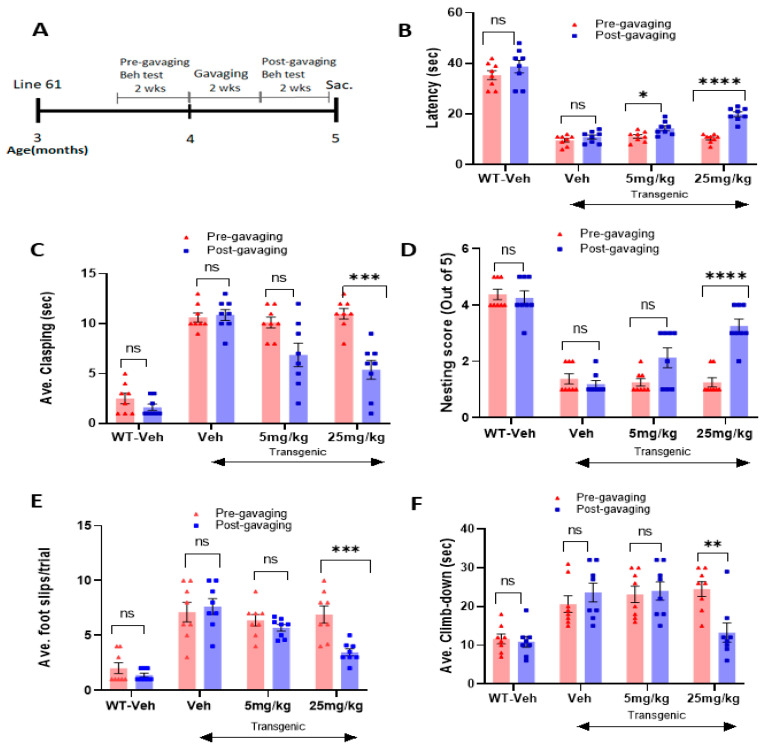
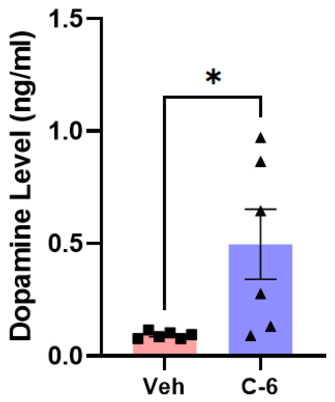
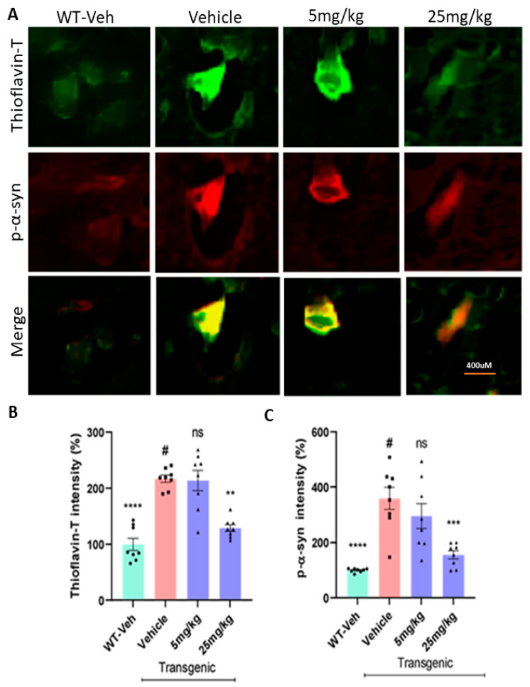
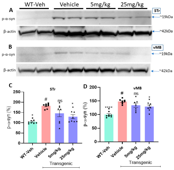
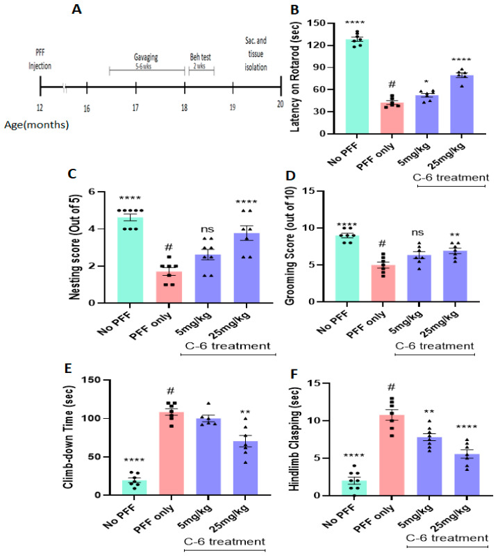
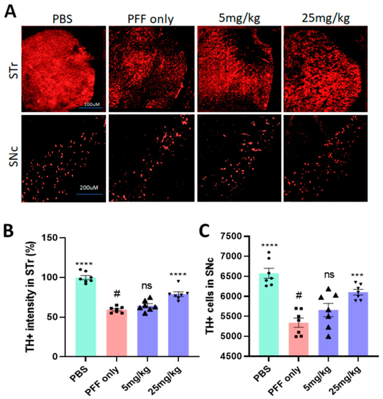
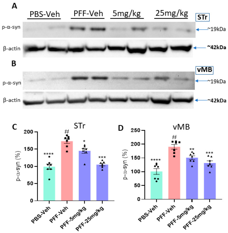
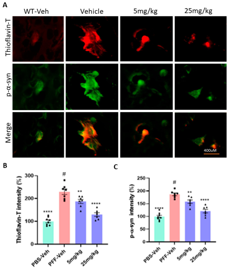
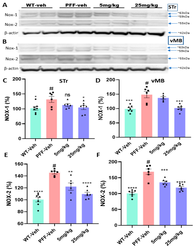
Parkinson's disease (PD) is a progressive neurodegenerative motor disorder without an available therapeutic to halt the formation of Lewy bodies for preventing dopaminergic neuronal loss in the nigrostriatal pathway. Since oxidative-stress-mediated damage has been commonly reported as one of the main pathological mechanisms in PD, we assessed the efficacy of a novel NOX inhibitor from AptaBio Therapeutics (C-6) in dopaminergic cells and PD mouse models. The compound reduced the cytotoxicity and enhanced the cell viability at various concentrations against MPP+ and α-synuclein preformed fibrils (PFFs). Further, the levels of ROS and protein aggregation were significantly reduced at the optimal concentration (1 µM). Using two different mouse models, we gavaged C-6 at two different doses to the PD sign-displaying transgenic mice for 2 weeks and stereotaxically PFF-injected mice for 5 weeks. Our results demonstrated that both C-6-treated mouse models showed alleviated motor deficits in pole test, hindlimb clasping, crossbeam, rotarod, grooming, and nesting analyses. We also confirmed that the compound treatment reduced the levels of protein aggregation, along with phosphorylated-α-synuclein, in the striatum and ventral midbrain and further dopaminergic neuronal loss. Taken together, our results strongly suggest that NOX inhibition can be a potential therapeutic target for PD.

Keywords: ROS inhibition; Thioflavin T; oxidative stress; protein aggregation; α-synuclein preformed fibrils.


Conflict of interest statement

This study was performed in collaboration between the Kim Lab at Delaware State University and AptaBio Therapeutics Inc. The novel compound was designed and synthesized by AptaBio Therapeutics, which partially supported this collaborative project. However, AptaBio Inc had no role in the experimental designs, data collection, analyses, and interpretation, or the writing of the manuscript for publication.

Figures

Figure 1
Figure 1
The treatment of a novel NOX inhibitor increases cell viability and decreases cytotoxicity derived from MPP+ or PFF in N27P dopaminergic cells. The optimal dose of the compound-6 was screened at various concentrations (1 nM–10 µM) in MTT (A,C) and LDH assay (B,D). The most effective concentration of the C-6 was consistently 1 µM against 640 µM MPP+ (A,B) and PFF (C,D) in both assays. (A,B) The C-6 increased cell viability and reduced the level of LDH (cytotoxicity) derived from MPP+ at the optimal dose of 1 µM. (C,D) The C-6 treatment increased the cell viability and decreased the cytotoxicity (LDH level) induced by PFF at the concentration between 100 nM and 1 µM. One-way ANOVA with Dunnett’s test was applied for statistical significance, compared with MPP+ only (#) or PFF only control (#) (n = 4/group independently). The no MPP+ (or no PFF) group is considered as positive control and converted to 100% for showing relativity. *: p < 0.05; **: p < 0.01; ***: p < 0.001, ****: p < 0.0001 and ns: not significant.
Figure 2
Figure 2
The compound-6 reduces the level of ROS generated by MPP+-induced toxicity in N27 cells. (A) The example images show that MPP+ exposure triggered a significant increase in ROS generation, which was reversed by the compound treatment, compared with no-MPP+ group. (B) The C-6 treatment (1 µM) markedly decreased the MPP+-induced ROS generation. One-way ANOVA with Dunnett’s test was adopted to compare with MPP+ only control (#) (n = 2–3 wells/experiment for 3 independent trials). ****: p < 0.0001.
Figure 3
Figure 3
The C-6 effectively alleviates PFF-induced ROS generation and protein aggregation. (A) The example images support that PFF-induced ROS generation and protein aggregates were substantially reversed by the C-6 treatment, compared with no PFF. (B,C) The C-6 treatment significantly ameliorated both protein aggregation and ROS generation, induced by PFF exposure. The optimal concentration used was 1 µM, at which ROS generation and protein aggregation were significantly reduced with the C-6. One-way ANOVA with Dunnett’s test was applied to compare the effect of the treatment with PFF only (#) (n = 3 or 4 independently). ****: p < 0.0001.
Figure 4
Figure 4
The oral treatment of the compound-6 for 2 weeks alleviates PD-like motor deficits in the L-61 transgenic mouse model. (A) the schematic diagram shows the gavaging and behavioral test schedule in timeline after transgenic mice turned 3 months old. The oral treatment improved the latency on rotarod (B), alleviated hindlimb clasping (C), enhanced nesting capabilities (D), reduced the slips on the crossbeam (E), and decreased the climb-down time on the pole test (F). Student’s paired t-test was performed to assess the effects of the compound treatment. Before treatment was labeled as pre-gavaging and after treatment as post-gavaging (n = 7–8/group). *: p < 0.05, **: p < 0.01, ***: p < 0.001 and ****: p < 0.0001. ns: not significant.
Figure 5
Figure 5
Two weeks of the compound-6 gavaging significantly increases the level of dopamine in the striatum of the L-61 mice. The treatment of 25 mg/kg substantially increased the dopamine released into the striatum. Unpaired Student’s t-test was applied to compare the treatment and vehicle groups (n = 6/group). *: p < 0.05.
Figure 6
Figure 6
The C-6 treatment decreases the levels of protein aggregation and phosphorylated α-synuclein in the L-61 mice. (A) A few example images show the different levels of protein aggregates in Thioflavin-T stain and phosphorylated Ser-129-α-synuclein in the striatum. The oral treatment reduced the levels of protein aggregates in Thioflavin-T (B) and phosphorylated Ser-129-α-synuclein (C) in the striatum of transgenic mice. One-way ANOVA with Dunnett’s test to compare the intensities of Thioflavin-T stain and phospho-Ser129-α-synuclein label present across the groups with or without treatment (#) (n = 7–8/group). **: p < 0.01, ***: p < 0.001 and ****: p < 0.0001. ns: not significant.
Figure 7
Figure 7
The compound treatment reduces the levels of phosphorylated Ser-129 α-synuclein in the striatum and the ventral midbrain of L-61 mice. (A,B) In Western blots, the oral treatment of the C-6 lowered the level of phosphorylated Ser-129 α-synuclein in the STr and vMB in a dose-dependent manner. The band intensity was also compared with vehicle-treated WT mice (WT-veh). (C,D) The oral C-6 treatment decreased the levels of phosphorylated Ser-129-α-synuclein in the striatum (C) and the ventral midbrain (vMB) (D). One-way ANOVA with Dunnett’s test was used to compare the band intensities of phospho-Ser129-α-synuclein among the treatments, vehicles (#), and vehicle-treated WT (n = 7–8/group). *: p < 0.05 and ****: p < 0.0001. ns: not significant.
Figure 8
Figure 8
The compound-6 treatment alleviates motor dysfunctions in PFF-injected mice. (A) The schematic diagram shows the timeline of gavaging and behavioral test scheduled in PFF-injected mice. The oral treatment for 5–6 weeks improved the latency on rotarod (B), enhanced nesting capabilities (C), improved grooming (D), reduced the climb-down time on the pole test (E), and alleviated hindlimb clasping (F). One-way ANOVA with Dunnett’s test was applied to compare the treated groups with that of PFF-only mice (#) (n = 7–8/group). *: p < 0.05, **: p < 0.01 and ****: p < 0.0001. ns: not significant.
Figure 9
Figure 9
The C-6 treatment prevents or even recovers from dopaminergic neuronal damage in the SNc and increases TH intensity in the STr. (A) The examples of IHC show an increase in TH intensity in the STr and higher number of TH+ neurons in the SNc from mice treated with 25 mg/kg dose, compared with PFF only. (B) The C-6 treatment at 25 mg/kg enhanced the level of TH+ in the striatum, compared with PFF only (n = 7–8/group). (C) The number of TH+ neurons in the SNc is significantly higher with the C-6 treatment than PFF only (n = 7–8/group). One-way ANOVA with Dunnett’s test was used to compare the intensity of TH+ cells in the striatum and TH+ cell number in the SNc, compared with PBS-vehicle- or PFF-vehicle (#)-treated mice. ***: p < 0.001 and ****: p < 0.0001. ns: not significant.
Figure 10
Figure 10
The C-6 treatment reduces the level of phosphorylated Ser-129-α-synuclein in the striatum and the ventral midbrain in PFF-injected mice. (A,B) As revealed by Western blots, PFF injection increased the level of phosphorylated Ser-129-α-synuclein, whereas the compound reduced the level in STr (A) and vMB (B). (C,D) The oral treatment of the compound-6 at both 5 mg/kg and 25 mg/kg significantly reduced the levels of phosphorylated Ser-129-α-synuclein in the striatum (C) as well as in the vMB (D) in a dose-dependent manner. One-way ANOVA with Dunnett’s test was applied to compare the expression level of phospho-Ser129-α-synuclein among the groups with or without treatment (#) (n = 7–8/group). *: p < 0.05, **: p < 0.01, ***: p < 0.001 and ****: p < 0.0001.
Figure 11
Figure 11
In the immunohistochemical analyses, the C-6 treatment decreases the levels of protein aggregates and phosphorylated Ser-129-α-synuclein in the striatum of PFF-injected mice. (A) Based on fluorescent co-labeling in immunohistochemistry, PFF injection enhanced the levels of protein aggregates in Thioflavin-T stain and phosphorylated Ser-129-α-synuclein, whereas the C-6 treatment substantially reduced both labeling intensities in the striatum. (B,C) In the analysis of Thioflavin-T label intensity, both 5 mg/kg and 25 mg/kg treatments significantly reduced the label intensity of protein aggregates (B) and phosphorylated Ser-129-α-synuclein (C) in the striatum. One-way ANOVA with Dunnett’s test was adopted to compare the intensity of Thioflavin-T stain and phospho-Ser129-α-synuclein label present across the groups with or without treatment (#) (n = 7–8/group). **: p < 0.01 and ****: p < 0.0001.
Figure 12
Figure 12
The C-6 treatment reduces the expression levels of NOX-1 and NOX-2 in the STr and vMB in Western blot analyses. (A,B) The examples of WB images show that both NOX-1 and -2 were upregulated by PFF injection, while the C-6 treatment reduced both levels in the STr (A) and vMB (B) in a dose-dependent manner. (C,D) The elevated NOX-1 level by PFF injection was reduced by the compound treatment in the STr (C) and vMB (D). (E,F) The PFF injection significantly increased the level of NOX-2, which was reversed by the C-6 treatment in the STr (E) and vMB (F). One-way ANOVA with Dunnett’s test was adopted to compare the expressions of NOX-1 or -2 across the groups with or without treatment (#), compared to WT-Veh (n = 7–8/group). *: p < 0.05, **: p < 0.01, ***: p < 0.001 and ****: p < 0.0001. ns: not significant.

References

    1. Spillantini M.G., Schmidt M.L., Lee V.M.-Y., Trojanowski J.Q., Jakes R., Goedert M. α-Synuclein in Lewy bodies. Nature. 1997;388:839–840. doi: 10.1038/42166. - DOI - PubMed
    1. Braak H., del Tredici K. Neuroanatomy and Pathology of Sporadic Parkinson’s Disease. Adv. Anat. Embryol. Cell Biol. 2009;201:1–119. - PubMed
    1. Siderowf A., Stern M. Update on Parkinson Disease. Ann. Intern. Med. 2003;138:651. doi: 10.7326/0003-4819-138-8-200304150-00013. - DOI - PubMed
    1. Stewart T., Sossi V., Aasly J.O., Wszolek Z.K., Uitti R.J., Hasegawa K., Yokoyama T., Zabetian C.P., Leverenz J.B., Stoessl A.J., et al. Phosphorylated α-synuclein in Parkinson’s disease: Correlation depends on disease severity. Acta Neuropathol. Commun. 2015;3:7. doi: 10.1186/s40478-015-0185-3. - DOI - PMC - PubMed
    1. Belarbi K., Cuvelier E., Destée A., Gressier B., Chartier-Harlin M.-C. NADPH oxidases in Parkinson’s disease: A systematic review. Mol. Neurodegener. 2017;12:84. doi: 10.1186/s13024-017-0225-5. - DOI - PMC - PubMed