. 2022 Apr 14;23(8):4348.
doi: 10.3390/ijms23084348.

Amylin Protein Expression in the Rat Brain and Neuro-2a Cells

Affiliations

Amylin Protein Expression in the Rat Brain and Neuro-2a Cells

Yeong-Min Yoo et al. Int J Mol Sci. .

Abstract

The localization and expression of amylin protein in the rodent brain and mouse neuroblastoma Neuro-2a (N2a) are less widely known. Thus, this study investigated the expression distribution of amylin in the rat brain and N2a treated with steroid hormones. Amylin protein was identified in the olfactory bulb, cerebral cortex, dentate gyrus, thalamus, hypothalamus, ventral tegmental area (VTA), cerebellum, and brain stem in the rat brain. Additionally, the amylin protein was localized with the mature neurons of the cerebral cortex and dopaminergic neurons of the VTA. Progesterone (P4) and dexamethasone (Dex) significantly decreased, and 17β-estradiol (E2) increased the amylin protein level in the cerebral cortex. The P4 receptor antagonist RU486 significantly influenced the effects of P4 and Dex, and the E2 receptor antagonist ICI 182,780 slightly changed E2's effect. Amylin protein expression was significantly reduced in the VTA by P4 and Dex, and its expression was changed only following P4 plus RU486 treatment. It was confirmed for the first time that amylin protein is strongly expressed in the cytoplasm in N2a cells using immunofluorescent staining. P4 increased the levels of amylin, and RU486 treatment decreased them. Dex significantly increased the levels of amylin protein. RU486 treatment reversed the effects of Dex. Therefore, amylin protein is expressed in the cerebral cortex neurons and dopaminergic neurons of the VTA of the immature rat brain. P4 and Dex influence the expression of amylin protein in the rat brain and N2a cells.

Keywords: 17β-estradiol; Neuro-2a; amylin; dexamethasone; progesterone; rat brain.


Conflict of interest statement

The authors declare no conflict of interest.

Figures

Figure 1
Figure 1
Distribution of amylin in the immature rat brain. Samples from different regions of the olfactory bulb, cerebral cortex, dentate gyrus, thalamus, hypothalamus, ventral tegmental area, cerebellum, and brain stem were analyzed in immature rat brains. Fluorescence photomicrographs showed amylin in a sagittal section. Amylin is expressed widely in the brain. Scale bar, 50 μm.
Figure 2
Figure 2
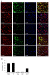
Colocalization of amylin protein with neuron-specific markers in the immature rat brain. (A) Amylin protein was co-expressed with neuron-specific markers such as NeuN, a marker of mature neurons; TH, a marker of dopaminergic neurons; IBA1, a marker of microglia; olig2, a marker of oligodendrocytes; and GFAP, a marker of astrocytes. Amylin protein was colocalized with mature neurons in the cerebral cortex and dopaminergic neurons in the VTA. (B) Quantification of colocalization in the immature rat brain. Colocalization was shown in yellow in the merged image. Colocalization analysis was performed using ImageJ software. Scale bar, 50 μm.
Figure 3
Figure 3
Amylin protein expression changes induced by steroid hormones in immature rat brain regions. Immature SD rats (postnatal days 10–14) were subcutaneously injected with 17β-estradiol (E2, 50 μg/kg body weight (BW)), dexamethasone (Dex, 10 mg/kg BW), progesterone (P4, 20 mg/kg BW), or vehicle (corn oil; Sigma-Aldrich) once per day for 5 consecutive days. ICI 182,780 (10 mg/kg BW) and mifepristone (RU486, 50 mg/kg BW) were treated 30 min before hormone administration. Amylin protein was then detected by Western blotting. The relative amounts of proteins were quantified as described in Section 4. Data are presented as the mean ± standard error of three experiments. * p < 0.05, ** p < 0.01, *** p < 0.001 vs. the control of cerebral cortex; ++ p < 0.01, +++ p < 0.001 vs. the control of the VTA; %% p < 0.01, E2 vs. E2 + ICI; ## p < 0.01 and ### p < 0.001, P4 vs. P4 + RU486; & p < 0.05 and && p < 0.01, Dex vs. Dex + RU486. Ve, vehicle; E2, 17β-estradiol; P4, progesterone, Dex dexamethasone; I, ICI 182,780; R, RU486.
Figure 4
Figure 4
Amylin protein expression in the cytoplasm of N2a cells (A). Box indicates the magnified area (B). Immunofluorescent staining was described in Materials and Methods. Scale bar, 20 μm.
Figure 5
Figure 5
Amylin protein and ERK expression changes induced by P4 treatment in N2a cells. N2a cells were cultured in DMEM with 10% FBS, 100 µg/mL streptomycin sulfate, and 100 U/mL penicillin G sodium at 37 °C and 5% CO2. Cells treated with P4 (10−7, 10−8 M) with/without antagonist (RU486, 10−6 M) for 24 h. N2a cell morphology (A). Cell viability (B). Western blotting detected amylin, p-ERK, and progesterone receptor (PR) (C). The relative amounts of proteins were quantified as described in Section 4. Data were presented as the mean ± standard error of three experiments. * p < 0.05 and *** p < 0.001 vs. the control; ### p < 0.001 and &&& p < 0.001, P4 vs. P4 + RU486. Scale bar, 25 μm.
Figure 6
Figure 6
Amylin protein and ERK expression changes induced by Dex treatment in N2a cells. N2a cells were cultured in DMEM with 10% FBS, 100 µg/mL streptomycin sulfate, and 100 U/mL penicillin G sodium at 37 °C and 5% CO2. Cells treated with Dex (10−7, 10−8 M) with/without antagonist (RU486, 10−6 M) for 24 h. N2a cell morphology (A). Cell viability (B). Western blotting detected amylin, p-ERK, and progesterone receptor (PR) (C). The relative amounts of proteins were quantified as described in Section 4. Data are presented as the mean ± standard error of three experiments. * p < 0.05, ** p < 0.01 and *** p < 0.001 vs. the control; ## p < 0.01, ### p < 0.001 and & p < 0.05, P4 vs. P4 + RU486. Scale bar, 25 μm.

Similar articles

Cited by

References

    1. Westermark P., Wernstedt C., Wilander E., Hayden D.W., O’Brien T.D., Johnson K.H. Amyloid fibrils in human insulinoma and islets of Langerhans of the diabetic cat are derived from a neuropeptide-like protein also present in normal islet cells. Proc. Natl. Acad. Sci. USA. 1987;84:3881–3885. doi: 10.1073/pnas.84.11.3881. - DOI - PMC - PubMed
    1. Cooper G.J., Willis A.C., Clark A., Turner R.C., Sim R.B., Reid K.B. Purification and characterization of a peptide from amyloid-rich pancreases of type 2 diabetic patients. Proc. Natl. Acad. Sci. USA. 1987;84:8628–8632. doi: 10.1073/pnas.84.23.8628. - DOI - PMC - PubMed
    1. Hay D.L., Chen S., Lutz T.A., Parkes D.G., Roth J.D. Amylin: Pharmacology, Physiology, and Clinical Potential. Pharmacol. Rev. 2015;67:564–600. doi: 10.1124/pr.115.010629. - DOI - PubMed
    1. Lutz T.A. Control of energy homeostasis by amylin. Cell. Mol. Life Sci. 2012;69:1947–1965. doi: 10.1007/s00018-011-0905-1. - DOI - PMC - PubMed
    1. Wookey P.J., Lutz T.A., Andrikopoulos S. Amylin in the periphery II: An updated mini-review. Sci. World J. 2006;6:1642–1655. doi: 10.1100/tsw.2006.263. - DOI - PMC - PubMed

LinkOut - more resources