. 2022 Apr 16;23(8):4420.
doi: 10.3390/ijms23084420.

Anti-Inflammatory and Neuroprotective Mechanisms of GTS-21, an α7 Nicotinic Acetylcholine Receptor Agonist, in Neuroinflammation and Parkinson's Disease Mouse Models

Affiliations

Anti-Inflammatory and Neuroprotective Mechanisms of GTS-21, an α7 Nicotinic Acetylcholine Receptor Agonist, in Neuroinflammation and Parkinson's Disease Mouse Models

Jung-Eun Park et al. Int J Mol Sci. .

Abstract

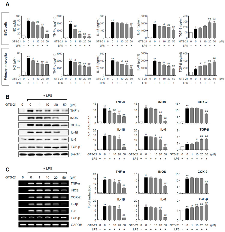
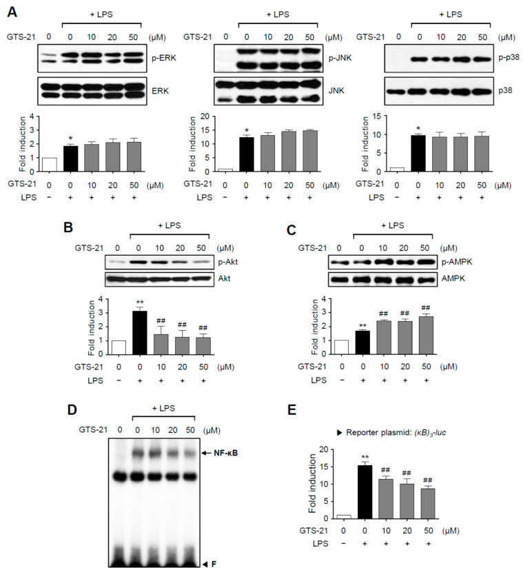
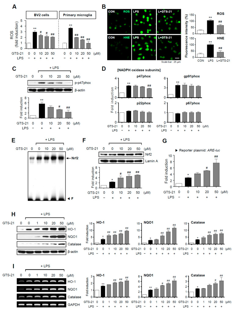
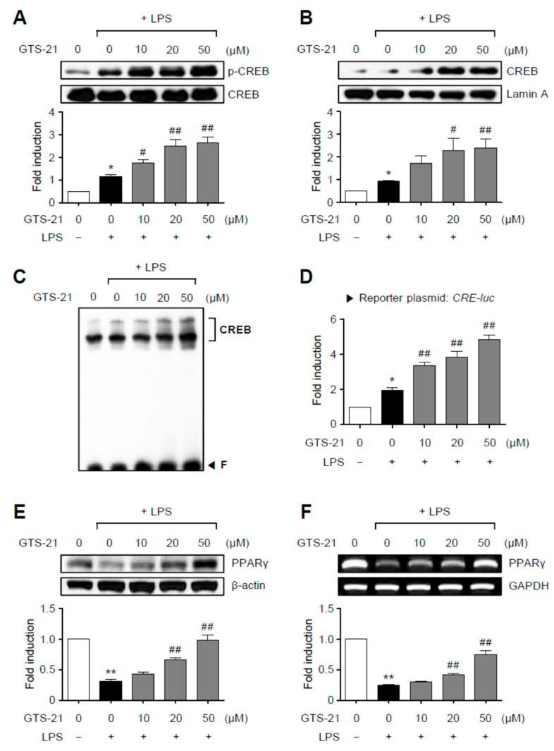
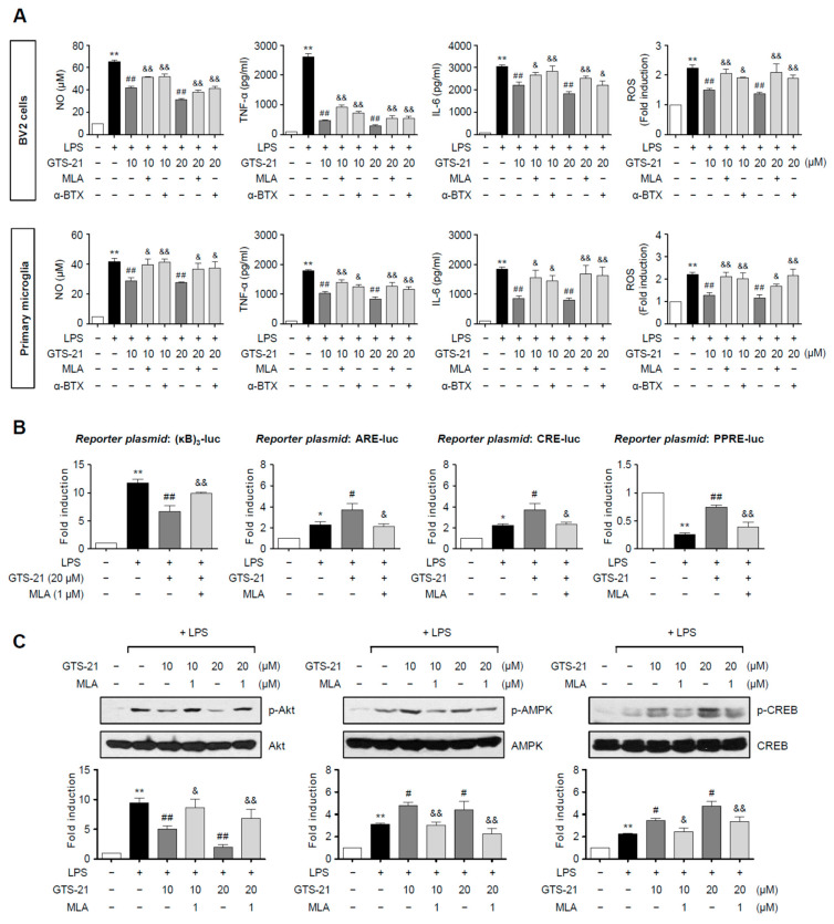
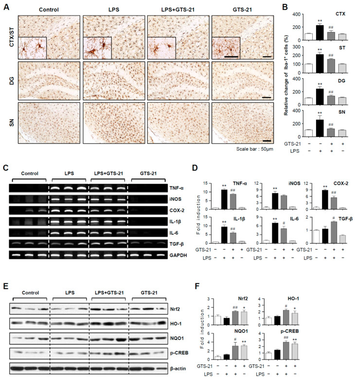
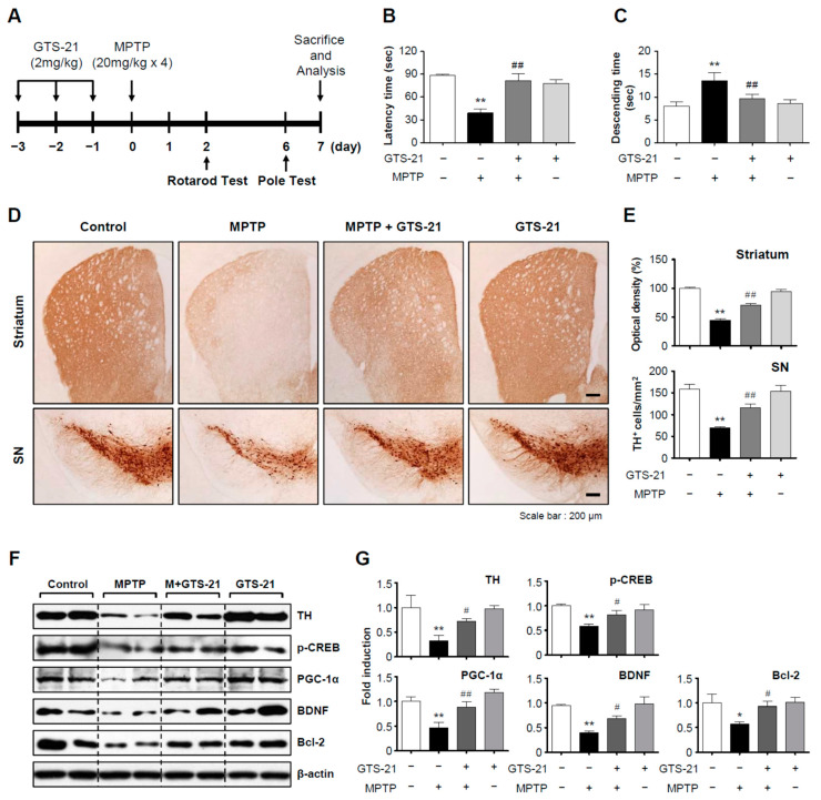
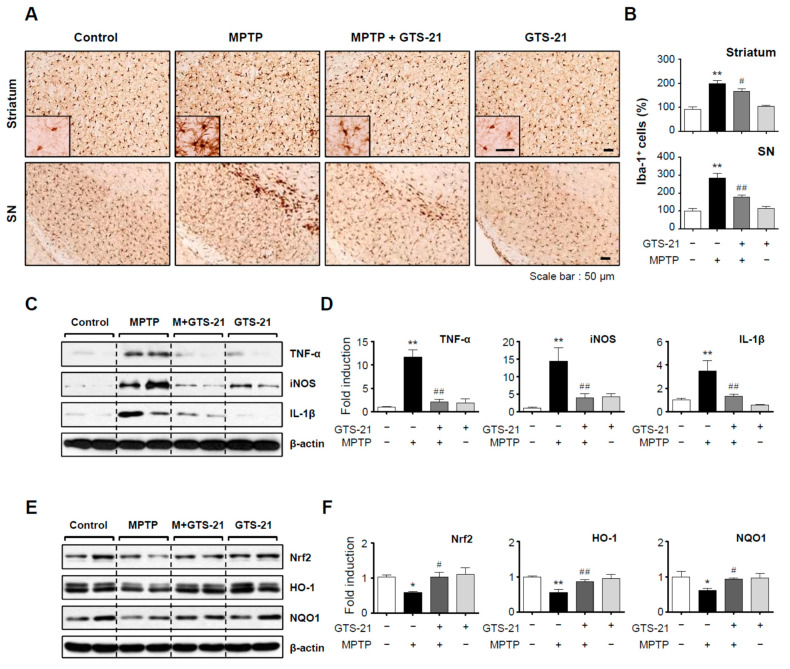
Neuroinflammation is crucial in the progression of neurodegenerative diseases. Thus, controlling neuroinflammation has been proposed as an important therapeutic strategy for neurodegenerative disease. In the present study, we examined the anti-inflammatory and neuroprotective effects of GTS-21, a selective α7 nicotinic acetylcholine receptor (α7 nAChR) agonist, in neuroinflammation and Parkinson's disease (PD) mouse models. GTS-21 inhibited the expression of inducible nitric oxide synthase (iNOS) and proinflammatory cytokines in lipopolysaccharide (LPS)-stimulated BV2 microglial cells and primary microglia. Further research revealed that GTS-21 has anti-inflammatory properties by inhibiting PI3K/Akt, NF-κB, and upregulating AMPK, Nrf2, CREB, and PPARγ signals. The effects of GTS-21 on these pro-/anti-inflammatory signaling molecules were reversed by treatment with an α7 nAChR antagonist, suggesting that the anti-inflammatory effects of GTS-21 are mediated through α7 nAChR activation. The anti-inflammatory and neuroprotective properties of GTS-21 were then confirmed in LPS-induced systemic inflammation and MPTP-induced PD model mice. In LPS-injected mouse brains, GTS-21 reduced microglial activation and production of proinflammatory markers. Furthermore, in the brains of MPTP-injected mice, GTS-21 restored locomotor activity and dopaminergic neuronal cell death while inhibiting microglial activation and pro-inflammatory gene expression. These findings suggest that GTS-21 has therapeutic potential in neuroinflammatory and neurodegenerative diseases such as PD.

Keywords: GTS-21; Parkinson’s disease; microglia; molecular mechanism; neuroinflammation; α7 nAChR agonist.


Conflict of interest statement

The authors declare no conflict of interest.

Figures

Figure 1
Figure 1
Effect of GTS-21 on inflammatory molecules in LPS-stimulated microglial cells. (A) After pretreatment with GTS-21 for 1 h, BV2 cells or primary cultured microglia were incubated with LPS (100 ng/mL for BV2, 10 ng/mL for primary microglia). The levels of nitrite, TNF-α, IL-1β, IL-6, and TGF-β were assessed in supernatants after a 16 h incubation period (n = 3–5 per group). (B,C) BV2 cells were treated with GTS-21 for 1 h before being incubated with LPS for 6 h. Western blot analysis (B) and RT-PCR (C) were performed to determine the expression level of inflammatory molecules (n = 3–4 per group). The left panel shows representative blots/gels, whereas the right panel shows quantitative data. The data are presented as the mean ± SEM. * p < 0.05, vs. control group; ** p < 0.01, vs. control group; # p < 0.05 vs. LPS-treated group; ## p < 0.01 vs. LPS-treated group.
Figure 2
Figure 2
Effect of GTS-21 on the activities of MAPKs, Akt, AMPK, and NF-κB in LPS-stimulated BV2 cells. (AC) Cell lysates from BV2 cells treated with LPS for 30 min in the presence or absence of GTS-21 were prepared, and western blot analysis was performed to investigate the effect of GTS-21 on MAPKs. (A), Akt (B), and AMPK (C) activity (n = 3–5 per group). The bottom panels show the quantification data. Phosphorylated MAPKs, Akt, and AMPK levels were adjusted to total forms and expressed as fold changes compared to untreated control samples. (D) Nuclear extracts from BV2 cells treated with GTS-21 in the presence of LPS for 1 h were used for EMSA for NF-κB. ‘F’ stands for free probe. (E) Analysis of the [κB]3-luc reporter gene activity after transient transfection (n = 5). The data are presented as the mean ± SEM. * p < 0.05, vs. control group; ** p < 0.01, vs. control group; ## p < 0.01 vs. LPS-treated group.
Figure 3
Figure 3
GTS-21 inhibited the production of ROS and HNE by inhibiting the NADPH oxidase subunit p47phox and increasing Nrf2/ARE signaling. (A) GTS-21 was applied to microglial cells 1 h before LPS stimulation for 16 h, and intracellular ROS levels were measured using the DCF-DA method (n = 4). (B) A representative confocal image of CellROX-derived fluorescence generated by intracellular ROS, and immunofluorescence staining for HNE in BV2 cells (n = 4). (C) Phosphorylation of the p47phox subunit was determined using western blot analysis (n = 3). (D) Quantitative RT-PCR to determiner mRNA expression level of NADPH oxidase subunits in BV2 microglia (n = 3–4 per group). (E) EMSA to assess the DNA binding activity of Nrf2. (F) The nuclear translocation of Nrf2 was assessed by western blot analysis (n = 3). (G) ARE-luc reporter gene activity after transient transfection (n = 3). (H,I) Effects of GTS-21 on the protein and mRNA expressions of HO-1, NQO1, and catalase in BV2 cells (n = 3). Western blot (H) and RT-PCR (I) data are shown. The left panel shows representative blots/gels, whereas the right panel shows quantitative data. The data are presented as the mean ± SEM. * p < 0.05, vs. control group; ** p < 0.01, vs. control group; # p < 0.05 vs. LPS-treated group; ## p < 0.01 vs. LPS-treated group.
Figure 4
Figure 4
GTS-21 enhanced CREB and PPARγ signaling in LPS-stimulated BV2 cells. (A) BV2 cells were pretreated for 1 h with GTS-21 at the indicated concentrations before being stimulated with LPS for 30 min (n = 3). The levels of phospho- and total CREB were determined using western blot analysis on cell lysates. (B) CREB nuclear translocation was detected using a western blot (n = 4). Quantification data are shown at bottom. (C) Nuclear extracts from BV2 cells treated with GTS-21 in the presence of LPS for 1 h were used to perform EMSA for CREB. (D) CRE-luc reporter gene activity after transient transfection (n = 3). (E,F) BV2 cells were treated with GTS-21 for 1 h before being incubated with LPS for 6 h. To determine the effects of GTS-21 on the protein and mRNA expressions of PPARγ, western blot analysis (E) and RT-PCR (F) were performed (n = 3–4 per group). The upper panel shows representative blots/gels, while the bottom panel shows quantitative data. The data are presented as the mean ± SEM. * p < 0.05, vs. control group; ** p < 0.01, vs. control group; # p < 0.05 vs. LPS-treated group; ## p < 0.01 vs. LPS-treated group.
Figure 5
Figure 5
α7 nAChR antagonists reversed the anti-inflammatory effects of GTS-21 in LPS-stimulated microglia. (A) The effect of methyllycaconitine (MLA) and α-bungarotoxin (α-BTX) on the production of NO, TNF-α, IL-6, and ROS in LPS + GTS-21-treated BV2 cells and primary microglia (n = 3–4 per group). Cells were pre-treated with MLA (1 μM) or α-BTX (0.01 μM) for 1 h, and then GTS-21 (10, 20 μM) for 1 h, followed by the treatment with LPS (100 ng/mL for BV2, 10 ng/mL for primary microglia) for 16 h. The levels of NO, TNF-α, and IL-6 released into the medium, as well as intracellular ROS, were measured. (B) The effect of MLA on NF-κB, Nrf2, CREB, and PPARγ reporter gene activities (n = 3–4 per group). BV2 cells were transfected with the reporter plasmid and then treated with MLA (1 μM) for 1 h, followed by GTS-21 (20 μM) and LPS (100 ng/mL). Cells were harvested after 6 h of LPS treatment and the reporter gene assay was carried out. (C) BV2 cells were pre-treated with MLA (1 μM) for 1 h, and then GTS-21 (10, 20 μM) for 1 h, followed by the treatment with LPS for 30 min to determine p-Akt, p-AMPK, and p-CREB levels by western blot analyses (n = 3–4 per group). The data are presented as the mean ± SEM. * p < 0.05, vs. control group; ** p < 0.01, vs. control group; # p < 0.05 vs. LPS-treated group; ## p < 0.01 vs. LPS-treated group; & p < 0.05 vs. LPS + GTS-21-treated group; && p < 0.01 vs. LPS + GTS-21-treated group.
Figure 6
Figure 6
Effect of GTS-21 on microglial activation, inflammatory markers, and antioxidant signaling molecules in the brains of LPS-injected mice. (A,B) Iba-1 immunohistochemical staining and quantification of Iba-1-positive microglia 24 h after LPS injection (n = 4 per group, 3 sections/brain). GTS-21 (5 mg/kg) reduced microglial activation in the prefrontal cortex (CTX), striatum (ST), dentate gyrus of the hippocampus (DG), and substantia nigra (SN) of LPS-injected mice. Data quantification (B) and representative images (A) are shown. Scale bars, 50 μm. (C,D) Effects of GTS-21 on iNOS, COX-2, and cytokines mRNA levels in the cortices of LPS-injected mice (n = 3). The quantification data (D) and representative gels (C) are shown. (E,F) Western blot analysis was performed on protein extracts from the cortex of each group using Nrf2, p-CREB, HO-1, and NQO1 antibodies (n = 4–5 per group). Representative blots (E) and quantification data (F) are shown. The data are presented as the mean ± SEM. * p < 0.05, vs. control group; ** p < 0.01, vs. control group; # p < 0.05 vs. LPS-treated group; ## p < 0.01 vs. LPS-treated group.
Figure 7
Figure 7
GTS-21 ameliorated the impaired movement and dopaminergic neuronal cell death in the brains of MPTP-injected mice. (A) The experimental procedure is depicted schematically. Mice were given GTS-21 (2 mg/kg, i.p.) daily for three days prior to MPTP injection. Mice were sacrificed 7 days after MPTP injection for histological and biochemical analyses. (B,C) The rotarod and pole tests were carried out two and six days after the MPTP injection, respectively (n = 8–9 per group). (D,E) TH-positive neuronal cells in the substantia nigra and striatum (representative pictures) (D). The optical density of TH-positive fibers in the striatum and the number of TH-positive cells in the substantia nigra were measured for quantitative analysis (E) (n = 4 per group, 3 sections/brain). (F,G) Protein extracts from the substantia nigra of each group were analyzed using TH, p-CREB, PGC-1, BDNF, and Bcl2 antibodies (n = 4). The figures show representative blots (F) and quantification data (G). The data are presented as the mean ± SEM. * p < 0.05, vs. control group; ** p < 0.01, vs. control group; # p < 0.05 vs. MPTP-treated group; ## p < 0.01 vs. MPTP-treated group.
Figure 8
Figure 8
Effect of GTS-21 on microglial activation, inflammatory markers, and antioxidant enzyme expression in the brains of MPTP-injected mice. (A) Iba-1-positive microglial cells in the substantia nigra and striatum (representative images). (B) Quantitative analysis was performed by measuring the number of Iba-1-positive cells (n = 4 per group, 3 sections/brain). (CF) Protein extracts from the substantia nigra of each group were analyzed using TNF-α, iNOS, and IL-1β antibodies or Nrf2, HO-1, and NQO1 antibodies (n = 4). The figures show representative blots (C,E) and quantification data (D,F). The data are presented as the mean ± SEM. * p < 0.05, vs. control group; ** p < 0.01, vs. control group; # p < 0.05 vs. MPTP-treated group; ## p < 0.01 vs. MPTP-treated group.

References

    1. Chitnis T., Weiner H.L. CNS inflammation and neurodegeneration. J. Clin. Investig. 2012;127:3577–3587. doi: 10.1172/JCI90609. - DOI - PMC - PubMed
    1. Ransohoff R.M. How neuroinflammation contributes to neurodegeneration. Science. 2016;353:777–783. doi: 10.1126/science.aag2590. - DOI - PubMed
    1. Catalin B., Cupido A., Iancau M., Albu C.V., Kirchhoff F. Microglia: First responders in the central nervous system. Rom. J. Morphol. Embryol. 2013;54:467–472. - PubMed
    1. Hickman S., Izzy S., Sen P., Morsett L., Khoury J.E. Microglia in neurodegeneration. Nat. Neurosci. 2018;21:1359–1369. doi: 10.1038/s41593-018-0242-x. - DOI - PMC - PubMed
    1. Cianciulli A., Porro C., Calvello R., Trotta T., Lofrumento D.D., Panaro M.A. Microglia mediated neuroinflammation: Focus on PI3K modulation. Biomolecules. 2020;10:137. doi: 10.3390/biom10010137. - DOI - PMC - PubMed

MeSH terms