Review
. 2022 Apr 12;14(8):1611.
doi: 10.3390/nu14081611.

Actions of Retinoic Acid in the Pathophysiology of HIV Infection

Affiliations
Review

Actions of Retinoic Acid in the Pathophysiology of HIV Infection

Neil Sidell et al. Nutrients. .

Abstract

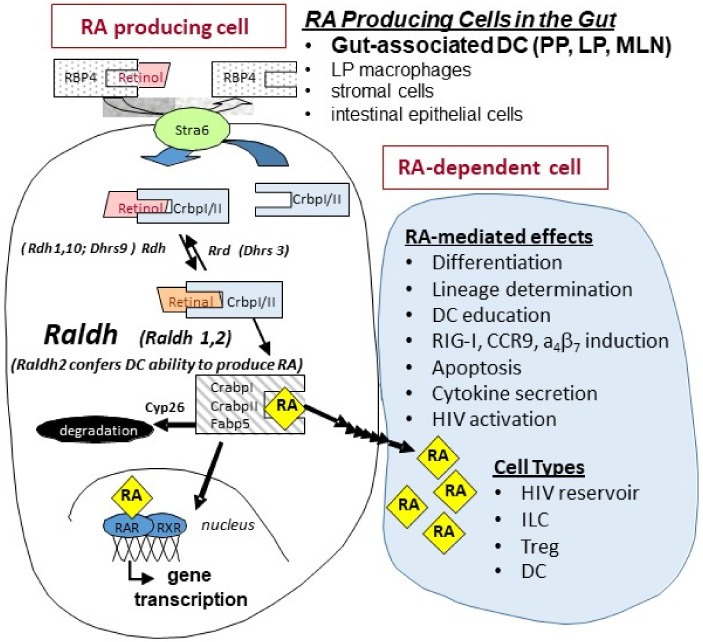
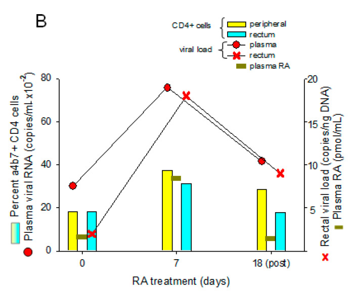
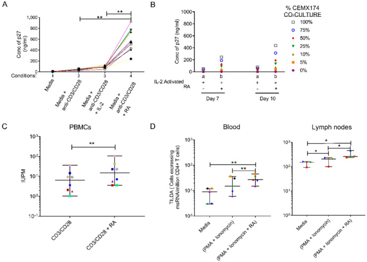
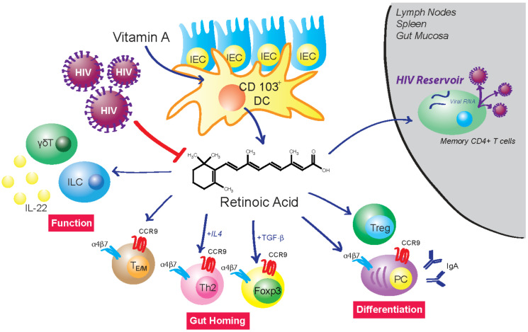
The vitamin A metabolite all-trans retinoic acid (RA) plays a key role in tissue homeostasis and mucosal immunity. RA is produced by gut-associated dendritic cells, which are among the first cells encountered by HIV. Acute HIV infection results in rapid reduction of RA levels and dysregulation of immune cell populations whose identities and function are largely controlled by RA. Here, we discuss the potential link between the roles played by RA in shaping intestinal immune responses and the manifestations and pathogenesis of HIV-associated enteropathy and similar conditions observed in SIV-infected non-human primate models. We also present data demonstrating the ability of RA to enhance the activation of replication-competent viral reservoirs from subjects on suppressive anti-retroviral therapy. The data suggest that retinoid supplementation may be a useful adjuvant for countering the pathologic condition of the gastro-intestinal tract associated with HIV infection and as part of a strategy for reactivating viral reservoirs as a means of depleting latent viral infection.

Keywords: HIV; immune regulation; mucosal immunity; retinoic acid; retinoids.


Conflict of interest statement

The authors declare no conflict of interest.

Figures

Figure 1
Figure 1
Retinoid metabolism and RA signaling. Studies in mice and humans have indicated that major cellular sources of all-trans retinoic acid (RA) production in the intestine are dendritic cells (DCs) from mesenteric lymph nodes (MLN), Peyer’s patches (PP), and small intestine lamina propria (LP); intestinal epithelial cells; and macrophages [1,2,12]. Raldh2 expression is restricted to limited cell types and is the critical regulatory element in immune cell populations that confers cells the ability to synthesize RA. Once formed, RA can be: (1) transported to the nucleus of the RA-producing cell (e.g., dendritic cells in the gut) where it binds to nuclear receptors and initiates gene transcription, (2) transported to neighboring cells (e.g., T-cells and ILCs) to initiate RA-mediated signaling, and/or (3) degraded by Cyp26.
Figure 2
Figure 2
GI mucosal damage results in loss of small intestine RA, which is reflected in reduced plasma RA levels. Loss of gut RA (jejunum, left) resulted in reduced plasma RA levels (right) in a partial body irradiation (PBI) with 5% bone marrow sparing (BM5) non-human primate (NHP) model of radiation-induced gut damage (rhesus macaques; 11.5 Gy PBI/BM5, n = 10; control, n = 10) as quantified by liquid chromatography-tandem mass spectrometry (LC-MS/MS); mean ± SEM (* p < 0.05). Adapted from Yu J, Huang W et al., 2021 [47].
Figure 3
Figure 3
Effects of acute SIV infection and anti-α4β7 antibody treatment on RA production and ILC3 frequency in NHP. NHP (Rhesus macaques) were infected with SIV at week 0 and treated with daily anti-retroviral therapy (ART) and control IgG (n = 7) (A) or ART and anti-α4β7 antibody (n = 8) (B) over the post-infection treatment periods indicated by the black arrows. (A) The ART/IgG regimen showed a rapid decrease in RA levels (blue) along with a decrease in the mean frequencies of ILC3 cells (red). (B) The ART/anti-α4β7 antibody regimen resulted in a recovery of plasma RA levels by week 15 (blue) equivalent to the pre-infection values that was accompanied by an expansion of the ILC3 cells (red). The ILC3 were obtained from the gated population of CD3−/CD8+/HLA-DR+/NKG2a−/NKp44+ cells in samples of colorectal tissue-isolated mononuclear cells from 3 representative animals from each group. RA levels (mean ± SEM) were quantified by LC-MS/MS and compared with pre-infection (week 0) values (* p < 0.05). Adapted from Byrareddy SN et al., 2016 [61].
Figure 4
Figure 4
Effects of SIV Infection on RA production in CD4+ T cells. Hut78 cells (CD4+ T cells) or chronically SIV-infected Hut78 cells [86] were treated with 2 µM retinol (substrate for RA synthesis) under serum free conditions for 4 h [87]. RA levels in cell pellets were quantified by LC-MS/MS [88]. SIV infection reduced the ability of Hut78 cells to make RA. * p < 0.05.
Figure 5
Figure 5
SIV-infected NHP and HIV-infected humans have similar decreases in plasma RA levels, which correlate with decreases in plasma RA levels observed in IBD relapse as compared to IBD remission. NHP (SIV+/−) and human (HIV+/−) plasma RA levels were quantified via LC-MS/MS, mean ± SEM. SIV (−), n = 10; SIV (+) 50 weeks, n = 5; HIV (−), n = 6; HIV (+) for ≥52 weeks, n = 6. IBD remission and IBD relapse levels were median plasma RA levels observed in n = 38 and n = 24 patients, respectively from a study by Paul et al. 2018 [85] (* p < 0.05). Adapted from Byrareddy SN et al., 2016 [61].
Figure 6
Figure 6
In vivo administration of RA to macaques with low viral loads. Monkeys were fed 10 mg/kg/day all-trans RA (orally) for 10 days, then followed for another 32 days. (A) Increased α4β7 expression on CD4+ lymphocytes. Central memory, effector memory, and naïve CD4+ lymphocytes were gated based on their dual expression of CD28 and CD95 [4] and quantified for percent α4β7+ cells. Naïve CD4+ T cells were categorized as CD28+/CD95, central memory CD4+ T cells as CD28+/CD95+, and effector memory CD4+ T cells as CD28/CD95+. Results represent the mean ± SEM of 4 monkeys. * p < 0.05, significant difference compared with baseline (day 0) values. (B) Representative example of a “spontaneous controller” monkey from “A” showing the correlation of plasma and rectal viral load with RA levels and expression of α4β7. Adapted from Olwenyi OA, 2020 [93].
Figure 6
Figure 6
In vivo administration of RA to macaques with low viral loads. Monkeys were fed 10 mg/kg/day all-trans RA (orally) for 10 days, then followed for another 32 days. (A) Increased α4β7 expression on CD4+ lymphocytes. Central memory, effector memory, and naïve CD4+ lymphocytes were gated based on their dual expression of CD28 and CD95 [4] and quantified for percent α4β7+ cells. Naïve CD4+ T cells were categorized as CD28+/CD95, central memory CD4+ T cells as CD28+/CD95+, and effector memory CD4+ T cells as CD28/CD95+. Results represent the mean ± SEM of 4 monkeys. * p < 0.05, significant difference compared with baseline (day 0) values. (B) Representative example of a “spontaneous controller” monkey from “A” showing the correlation of plasma and rectal viral load with RA levels and expression of α4β7. Adapted from Olwenyi OA, 2020 [93].
Figure 7
Figure 7
Retinoic Acid (RA) enhances viral replication in vitro and improves detection of the viral reservoir. (A) Levels of p27, a regulatory protein produced by HIV/SIV-infected cells [94], were measured in supernatant of peripheral blood mononuclear cells (PBMCs) from 10 randomly selected naive rhesus macaques (RMs). The PBMCs were infected with SIVmac239 after in vitro culturing with (1) media, unstimulated (2) anti-CD3/CD28 beads to activate the T-cell receptor (3) anti-CD3/CD28 + IL-2 (10 U/mL) and (4) anti-CD3/CD28 beads + IL-2 together with RA (1 μM) added under separate conditions on day 10. (B) Levels of p27 detected in supernatant fluids from pooled (n = 10 RMs) enriched CD4 T cells that were treated with either (a) anti-CD3/CD28 and IL-2 only or (b) anti-CD3/CD28 + IL-2 and RA. Thereafter, viral expansion was carried out by the co-culture with CEMX174 cells across different concentrations (0 to 100%) on days 7 and 10 respectively. (C) Quantitative Viral Outgrowth Assay (QVOA) of CD4+ T cells purified from PBMCs of anti-retroviral therapy (ART)-suppressed macaques indicating levels of Infectious Units per Million (IUPM) in CD3/CD28 versus CD3/CD28 + RA conditions (n = 8). (D) levels of msRNA transcripts obtained from enriched CD4 T cells collected from PBMC (n = 5 RMs) or auxiliary lymph node CD4+ T cells (n = 4 RMs) that were cultured in media only, media plus phorbol myristate acetate (PMA) + ionomycin and media plus PMA + ionomycin supplemented with retinoic acid. * shows p < 0.05 and ** represents p < 0.001 significant difference across studied groups obtained using Wilcoxon matched pairs signed rank tests. Reproduced with permission from Figure 1 from Olwenyi OA et al. [93].
Figure 8
Figure 8
HIV infection disrupts RA-mediated control of intestinal homeostasis. Mucosal CD103+ dendritic cells (DC) express Raldh2 and are a major source of RA production in the gut. They are among the first cells encountered by HIV and their infection inhibits RA biosynthesis. Studies have shown that acute HIV/SIV infection is closely linked to disruption of cell populations whose identity and function are regulated by RA. RA controls the differentiation of T cells into Tregs and B cells into IgA-producing plasma cells (PC). It also regulates the migration of lymphoid cells into the intestine by induction of gut-homing receptors α4β7 and CCR9. RA enhances IL-22 production from γδT cells and innate lymphoid cells (ILC). IL-22 plays a vital role in the regulation of multiple aspects of gut epithelial integrity, including control of epithelial cell growth and permeability, production of mucus and antimicrobial proteins, and complement production. RA supplementation can enhance activation of replication-competent HIV reservoirs. These data suggest the possible therapeutic use of retinoid derivatives to counteract HIV-associated enteropathy and as part of a “shock and kill” strategy to deplete latent viral infection.

Similar articles

Cited by

References

    1. Czarnewski P., Das S., Parigi S.M., Villablanca E.J. Retinoic Acid and Its Role in Modulating Intestinal Innate Immunity. Nutrients. 2017;9:68. doi: 10.3390/nu9010068. - DOI - PMC - PubMed
    1. Cassani B., Villablanca E.J., De Calisto J., Wang S., Mora J.R. Vitamin A and immune regulation: Role of retinoic acid in gut-associated dendritic cell education, immune protection and tolerance. Mol. Asp. Med. 2012;33:63–76. doi: 10.1016/j.mam.2011.11.001. - DOI - PMC - PubMed
    1. Larange A., Cheroutre H. Retinoic Acid and Retinoic Acid Receptors as Pleiotropic Modulators of the Immune System. Annu. Rev. Immunol. 2016;34:369–394. doi: 10.1146/annurev-immunol-041015-055427. - DOI - PubMed
    1. Iwata M., Hirakiyama A., Eshima Y., Kagechika H., Kato C., Song S.-Y. Retinoic Acid Imprints Gut-Homing Specificity on T Cells. Immunity. 2004;21:527–538. doi: 10.1016/j.immuni.2004.08.011. - DOI - PubMed
    1. Ross A.C. Vitamin A and retinoic acid in T cell–related immunity. Am. J. Clin. Nutr. 2012;96:1166S–1172S. doi: 10.3945/ajcn.112.034637. - DOI - PMC - PubMed