. 2022 Mar 25;14(4):678.
doi: 10.3390/v14040678.

New Insights into the Structure and Assembly of Bacteriophage P1

Affiliations

New Insights into the Structure and Assembly of Bacteriophage P1

Miguel F Gonzales et al. Viruses. .

Abstract

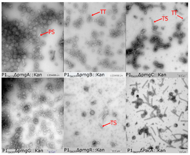
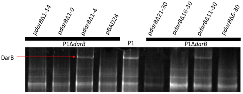
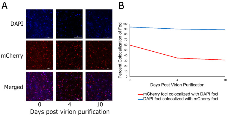
Bacteriophage P1 is the premier transducing phage of E. coli. Despite its prominence in advancing E. coli genetics, modern molecular techniques have not been applied to thoroughly understand P1 structure. Here, we report the proteome of the P1 virion as determined by liquid chromatography tandem mass-spectrometry. Additionally, a library of single-gene knockouts identified the following five previously unknown essential genes: pmgA, pmgB, pmgC, pmgG, and pmgR. In addition, proteolytic processing of the major capsid protein is a known feature of P1 morphogenesis, and we identified the processing site by N-terminal sequencing to be between E120 and S121, producing a 448-residue, 49.3 kDa mature peptide. Furthermore, the P1 defense against restriction (Dar) system consists of six known proteins that are incorporated into the virion during morphogenesis. The largest of these, DarB, is a 250 kDa protein that is believed to translocate into the cell during infection. DarB deletions indicated the presence of an N-terminal packaging signal, and the N-terminal 30 residues of DarB are shown to be sufficient for directing a heterologous reporter protein to the capsid. Taken together, the data expand on essential structural P1 proteins as well as introduces P1 as a nanomachine for cellular delivery.

Keywords: bacteriophage P1; bacteriophage structure; capsid targeting sequence; defense against restriction; proteomics; transmission electron microscopy.


Conflict of interest statement

The authors declare no conflict of interest.

Figures

Figure 1
Figure 1
Edman degradation of P1 major capsid protein gp23. (A) Edman degradation reveals the first six amino acids of the N-terminus of gp23 purified from P1 virions. (B) The amino acids identified in the open reading frame of gp23, in which the first 120 residues are processed from the gp23 N terminus.
Figure 2
Figure 2
Transmission electron micrographs of CsCl step-gradient purified P1 morphogenic gene knockouts and parental P1. PS is polysheath, TT is tail tube, and TS is truncated sheath.
Figure 3
Figure 3
The ability of DarB truncations to localize to the P1 capsid. Truncations of DarB residues 1–4 and 11–30 are tolerated, which would suggest amino acids 6 through 10 are required to localize DarB to the P1 virion. However, deletion of residues 16–30 and 21–30 also inhibit DarB packaging, suggesting that other deletions may induce improper folding of the N-terminus that inhibits recruitment of DarB to the P1 virion.
Figure 4
Figure 4
The 30 N-terminal residues of DarB are sufficient to target TetR-mCherry to the P1 capsid. (A) The simultaneous induction of the pBAD24_N30-tetR-mCherry expression vector and the P1ΔdarB lysogen results in the localization of TetR-mCherry to the P1 capsid. Fluorescence imaging of CsCl-purified P1ΔdarB + TetR-mCherry virions over days 0, 4, and 10 are shown using a DAPI filter, mCherry filter, and the two channels merged. (B) The line graph illustrates the colocalization of DAPI and mCherry foci over the 10 days post-purification of the P1ΔdarB + TetR-mCherry virions. The red line indicates the percent colocalization of mCherry foci with DAPI foci, and the blue line indicates the percent colocalization of DAPI foci with mCherry foci. This indicates that most (~90%) particles with packaged DNA contain TetR-mCherry, but many particles containing TetR-mCherry do not contain DNA; this may be due to particle instability or premature DNA ejection from the virions during storage.
Figure 5
Figure 5
The P1 genome and composition of the virion. (A): Genome map of phage P1 summarizing P1 morphogenesis genes. The size of the boxes indicates the size of each ORF, with genes encoded on the plus and minus strands staggered up and down, respectively. Genes colored black are involved in phage morphogenesis, based on the work presented here or in previous studies [10]. Products of genes outlined in red were detected in LC-MS/MS above the two-peptide threshold. Genes with bold labels were determined to be essential for morphogenesis by construction of single-gene deletions. The deletion of gray genes produced no antirestriction phenotype and no apparent defect in plating, thus these genes are considered non-essential with unknown function. Gene labels are color-coded to indicate their predicted location in the virion (panel B), with blue indicating the head, magenta the tail, and green the baseplate. (B): A schematic of the P1 virion, partitioned into three components of the head, tail, and baseplate (including tail fibers). Structural proteins are assigned to each area based on their predicted roles; color coding corresponds to the coding of gene labels in panel A. Sit is listed in both the tail and baseplate as its role as tape measure protein makes it a part of both components. Proteins PmgA, UpfC, gp5, and gp26 are marked with an asterisk as these are predicted to be part of the virion based on bioinformatics but were not detected by LC-MS/MS. Gene iddB omitted for simplicity.

References

    1. Bertani G. Studies on lysogenesis I: The mode of phage liberation by lysogenic Escherichia coli. J. Bacteriol. 1951;62:293–300. doi: 10.1128/jb.62.3.293-300.1951. - DOI - PMC - PubMed
    1. Lennox E. Transduction of linked genetic characters of the host by bacteriophage P1. Virology. 1955;1:190–206. doi: 10.1016/0042-6822(55)90016-7. - DOI - PubMed
    1. Yarmolinsky M.B., Sternberg N. Bacteriophage P1. In: Calendar R., editor. The Bacteriophages. Springer; Boston, MA, USA: 1988. pp. 291–438.
    1. Arber W., Dussoix D. Host specificity of DNA produced by Escherichia coli: I. Host controlled modification of bacteriophage λ. J. Mol. Biol. 1962;5:18–36. doi: 10.1016/S0022-2836(62)80058-8. - DOI - PubMed
    1. Iida S., Streiff M.B., Bickle T.A., Arber W. Two DNA antirestriction systems of bacteriophage P1, darA, and darB: Characterization of darA—Phages. Virology. 1987;157:156–166. doi: 10.1016/0042-6822(87)90324-2. - DOI - PubMed

Publication types

MeSH terms

LinkOut - more resources