. 2022 Apr 4;14(4):756.
doi: 10.3390/v14040756.

West Nile Virus Neuroinfection in Humans: Peripheral Biomarkers of Neuroinflammation and Neuronal Damage

Affiliations

West Nile Virus Neuroinfection in Humans: Peripheral Biomarkers of Neuroinflammation and Neuronal Damage

Orianne Constant et al. Viruses. .

Abstract

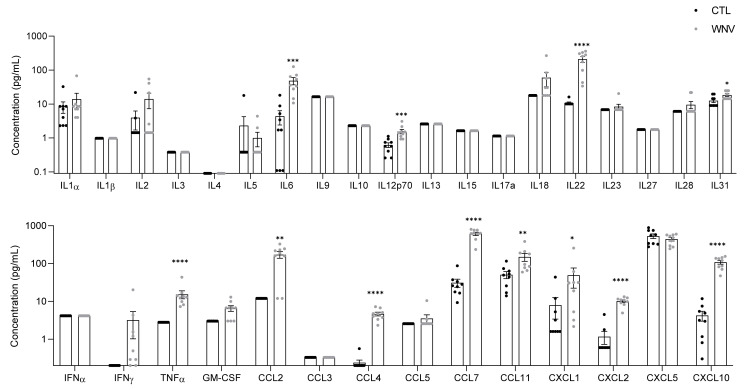
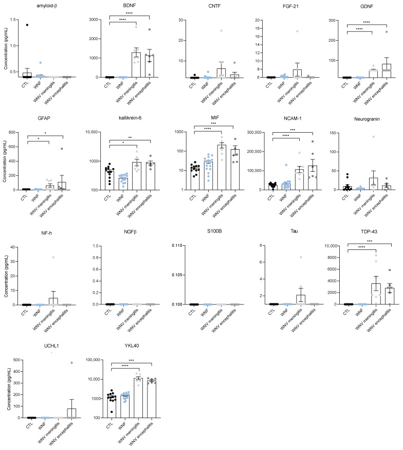
Among emerging arthropod-borne viruses (arbovirus), West Nile virus (WNV) is a flavivirus that can be associated with severe neuroinvasive infections in humans. In 2018, the European WNV epidemic resulted in over 2000 cases, representing the most important arboviral epidemic in the European continent. Characterization of inflammation and neuronal biomarkers released during WNV infection, especially in the context of neuronal impairments, could provide insight into the development of predictive tools that could be beneficial for patient outcomes. We first analyzed the inflammatory signature in the serum of WNV-infected mice and found increased concentrations of several inflammatory cytokines. We next analyzed serum and cerebrospinal-fluid (CSF) samples from a cohort of patients infected by WNV between 2018 and 2019 in Hungary to quantify a large panel of inflammatory cytokines and neurological factors. We found higher levels of inflammatory cytokines (e.g., IL4, IL6, and IL10) and neuronal factors (e.g., BDNF, GFAP, MIF, TDP-43) in the sera of WNV-infected patients with neuroinvasive disease. Furthermore, the serum inflammatory profile of these patients persisted for several weeks after initial infection, potentially leading to long-term sequelae and having a deleterious effect on brain neurovasculature. This work suggests that early signs of increased serum concentrations of inflammatory cytokines and neuronal factors could be a signature underlying the development of severe neurological impairments. Biomarkers could play an important role in patient monitoring to improve care and prevent undesirable outcomes.

Keywords: West Nile virus; flavivirus; neuroinvasive disease; peripheral biomarkers.


Conflict of interest statement

The authors declare no conflict of interest.

Figures

Figure A1
Figure A1
Biomarkers of inflammation in WNV-patients compared to healthy individuals. Plasma concentration in pg/mL of different pro-inflammatory markers of non-infected (black) and WNV-infected (grey) humans, measured by multiplex bead-based ELISA. Bars show means ± SEM (* p < 0.05, ** p < 0.01, *** p < 0.001, **** p < 0.0001).
Figure A2
Figure A2
Same inflammation profile in patients with meningitis, encephalitis, or meningoencephalitis. Plasma concentration in pg/mL of different pro-inflammatory markers of non-infected CTL (black), WNV-meningitis (light grey), WNV-encephalitis (dark grey) and WNV-meningoencephalitis (light red) humans, measured by multiplex bead-based ELISA. Bars show means ± SEM (* p < 0.05, ** p < 0.01, *** p < 0.001).
Figure A3
Figure A3
Examples of patients with persistent inflammation. Plasma concentration in pg/mL of different pro-inflammatory markers of non-infected CTL (black), WNND-short term (blue), and WNND-long term (grey) humans, measured by multiplex bead-based ELISA. Bars show value.
Figure 1
Figure 1
Upregulation of pro-inflammatory mediators after WNV infection in the serum of C57/BL6 mice (6 dpi). Plasma concentration in pg/mL of pro-inflammatory markers measured by multiplex bead-based ELISA in non-infected (black) and WNV-infected mice (grey). Bars show means ± SEM (* p < 0.05, ** p < 0.01, *** p < 0.001, **** p < 0.0001).
Figure 2
Figure 2
WNV infection results in systemic inflammation in patient sera. Plasma concentration in pg/mL of different pro-inflammatory markers of non-infected CTL (black), WNF (blue) and WNND (grey) humans, measured by multiplex bead-based ELISA. Bars show means ± SEM (* p < 0.05, ** p < 0.01, *** p < 0.001, **** p < 0.0001).
Figure 3
Figure 3
Identification of biomarkers of neuroinflammation in CSF of WNND patients. (A) CSF concentration in pg/mL of different neuronal markers of non-infected CTL (black), WNV-meningitis (yellow), and WNV-encephalitis (orange) humans, measured by multiplex bead-based ELISA. (B) IFNγ, GDNF, and neurogranin are differentially expressed in patients with meningitis and encephalitis. Bars show means ± SEM (* p < 0.05, ** p < 0.01).
Figure 4
Figure 4
Sera of WNND patients display markers of neuro-inflammation and neuronal injuries. Plasma concentration in pg/mL of different pro-inflammatory markers of non-infected CTL (black), WNF (blue), WNV-meningitis (light grey) and WNV-encephalitis (dark grey) humans, measured by multiplex bead-based ELISA. Bars show means ± SEM (* p < 0.05, ** p < 0.01, *** p < 0.001, **** p < 0.0001).
Figure 5
Figure 5
Persistence of inflammation in WNND patient sera. Plasma concentration in pg/mL of different pro-inflammatory markers of non-infected CTL (black), WNVF-short term (light blue), WNVF-long term (dark blue), WNVND-short term (light grey) and WNVND-long term (dark grey) humans, measured by multiplex bead-based ELISA. Bars show means ± SEM ((short term = 2 to 11 days, long term = more than 20 days); * p < 0.05 ** p < 0.01 *** p < 0.001).
Figure 6
Figure 6
WNND serum slightly perturbs the endothelium in a human BBB model. (A) Primary brain pericytes cultured in the basolateral compartment allow differentiation of CD34+ blood cord-derived endothelial cells in human brain-like endothelial cells cultured on transwell filters in the apical compartment. Serum can be added in the apical compartment of this human in vitro BBB model representing the blood. (B) Permeability coefficient (Pe) of the paracellular marker Lucifer Yellow was measured 4 days after the addition of CTL or WNND serum or DMSO as a positive control. ** p < 0.01.

References

    1. Kramer L.D., Li J., Shi P.Y. West Nile virus. Lancet Neurol. 2007;6:171–181. doi: 10.1016/S1474-4422(07)70030-3. - DOI - PubMed
    1. Campbell G.L., Marfin A.A., Lanciotti R.S., Gubler D.J. West Nile virus. Lancet Infect. Dis. 2002;2:519–529. doi: 10.1016/S1473-3099(02)00368-7. - DOI - PubMed
    1. Sejvar J.J., Marfin A.A. Manifestations of West Nile neuroinvasive disease. Rev. Med. Virol. 2006;16:209–224. doi: 10.1002/rmv.501. - DOI - PubMed
    1. Hayes E.B., Gubler D.J. West Nile virus: Epidemiology and clinical features of an emerging epidemic in the United States. Annu. Rev. Med. 2006;57:181–194. doi: 10.1146/annurev.med.57.121304.131418. - DOI - PubMed
    1. Calisher C.H., Gould E.A. Taxonomy of the virus family Flaviviridae. Adv. Virus Res. 2003;59:1–19. doi: 10.1016/S0065-3527(03)59001-7. - DOI - PubMed