. 2022 Apr 8;14(4):776.
doi: 10.3390/v14040776.

Brain Inflammation and Intracellular α-Synuclein Aggregates in Macaques after SARS-CoV-2 Infection

Affiliations

Brain Inflammation and Intracellular α-Synuclein Aggregates in Macaques after SARS-CoV-2 Infection

Ingrid H C H M Philippens et al. Viruses. .

Abstract

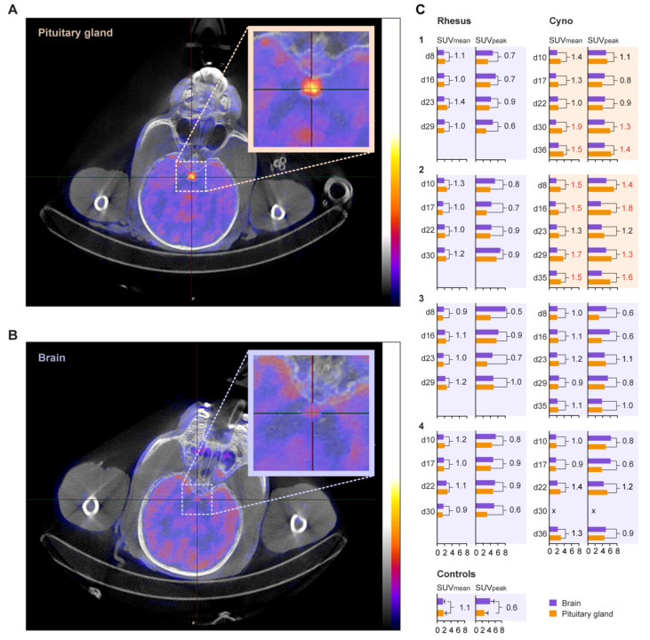
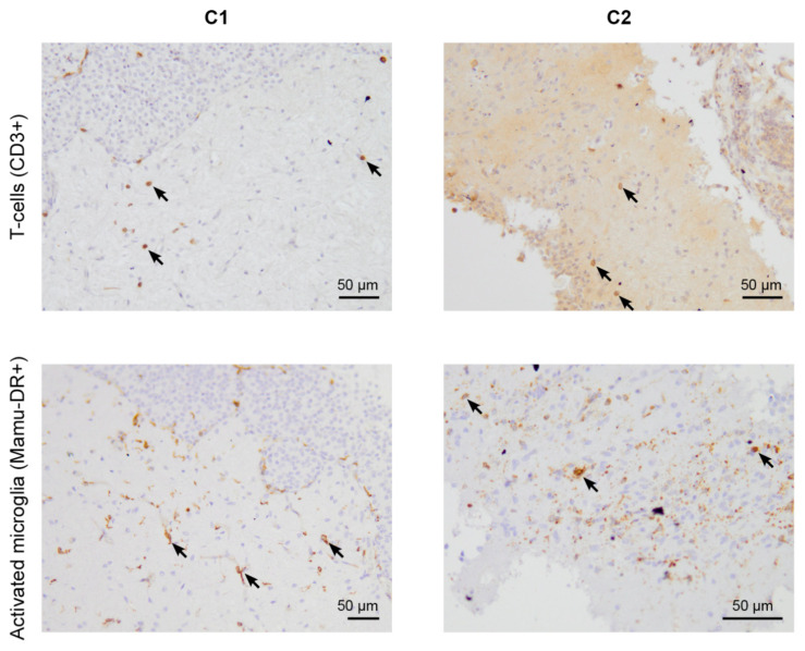
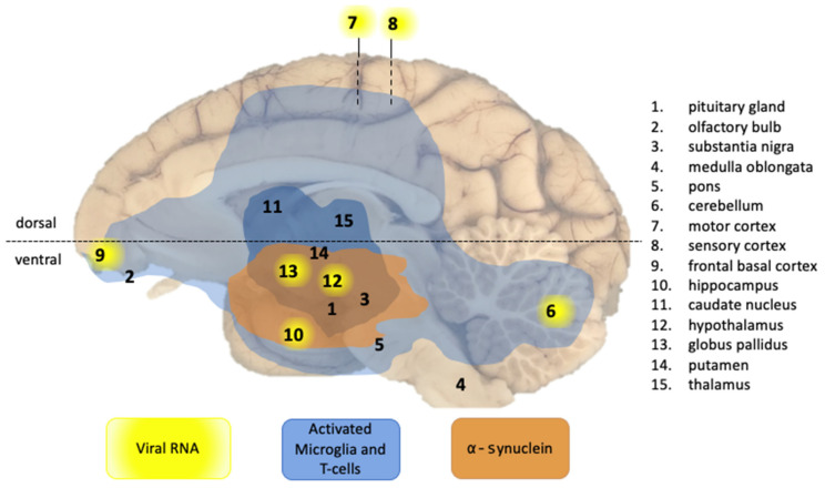
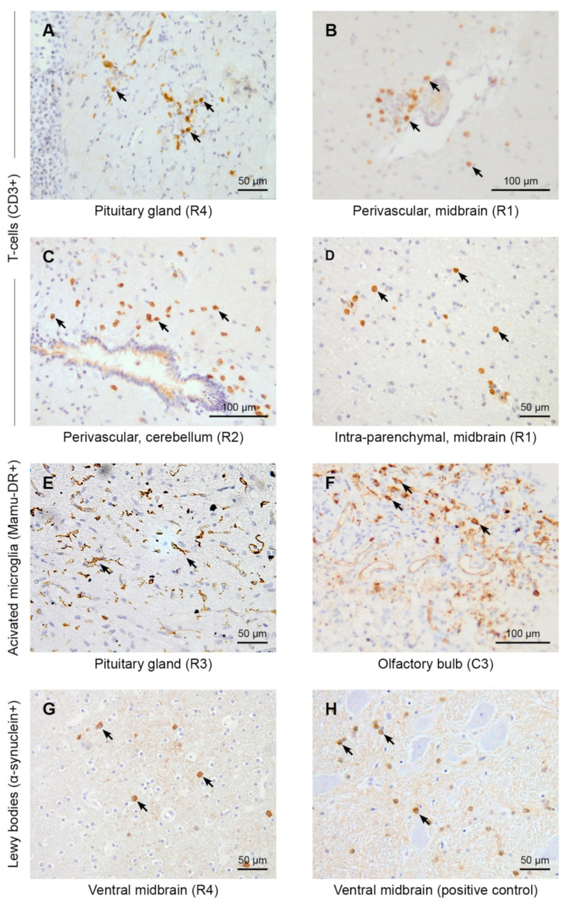
SARS-CoV-2 causes acute respiratory disease, but many patients also experience neurological complications. Neuropathological changes with pronounced neuroinflammation have been described in individuals after lethal COVID-19, as well as in the CSF of hospitalized patients with neurological complications. To assess whether neuropathological changes can occur after a SARS-CoV-2 infection, leading to mild-to-moderate disease, we investigated the brains of four rhesus and four cynomolgus macaques after pulmonary disease and without overt clinical symptoms. Postmortem analysis demonstrated the infiltration of T-cells and activated microglia in the parenchyma of all infected animals, even in the absence of viral antigen or RNA. Moreover, intracellular α-synuclein aggregates were found in the brains of both macaque species. The heterogeneity of these manifestations in the brains indicates the virus' neuropathological potential and should be considered a warning for long-term health risks, following SARS-CoV-2 infection.

Keywords: COVID-19; SARS-CoV-2; macaques; neuroinflammation; positron emission tomography-computed tomography; α-synuclein.


Conflict of interest statement

The authors declare no conflict of interest.

Figures

Figure 1
Figure 1
18F-FDG uptake in the pituitary gland after SARS-CoV-2 infection. Representative transversal slices of cynomolgus macaque C2, with increased 18F-FDG uptake in the pituitary gland, (A) and C3, with normal pituitary 18F-FDG uptake, (B) on day 8 are shown. The pituitary gland is indicated by the cross-hairs and boxed. Similar window-level settings are applied for both sections. (C) The standardized uptake values (SUV) of the brain and pituitary gland of each animal, per timepoint, are shown. On the Y-axis, the day post-infection of PET-CT and on the X-axis the SUV of the brain (purple bars) and pituitary gland (orange bars) are visualized for rhesus (left column) and cynomolgus (right column) macaques. For each animal, the SUVmean and SUVpeak are shown. The numbers in the graph represent the ratio of the SUV for the pituitary gland value divided by the SUV of the brain. A deviation from normal is defined as SUVmean ratio ≥ 1.5 and SUVpeak ratio ≥ 1. These ratios are indicated in red. Six non-infected animals were used as control for the definition of normal pituitary gland and brain SUVs. The animals boxed with an orange background showed increased uptake in the pituitary gland, while the animals boxed with a purple background do not show this increase.
Figure 2
Figure 2
Immunohistochemistry for T-cells and activated microglia (some positive cells are indicated with arrows) of SARS-CoV-2-infected brain sections of cynomolgus macaques C1 and C2. The presence of T-cells (CD3+) (top level) and activated microglia cells (Mamu-DR+) (bottom level) in the pituitary gland of monkeys C1 (left) and C2 (right), observed five weeks after infection.
Figure 3
Figure 3
Overview of CNS effects of SARS-CoV-2 infection detected in a macaque brain in this study. The presence of viral RNA was investigated in multiple regions of the brain, as indicated by the numbers. Viral RNA-positive regions in cynomolgus macaque C3 are indicated by a yellow background. Brain areas with infiltrated T-cells (CD3+) and activated microglia (Mamu-DR+) are shown in light blue (low expression) and dark blue (moderate expression). Brain areas with Lewy bodies (α-synuclein+) are depicted in orange. Blue and orange areas comprise of the combined observations from all animals. The horizontal dotted line indicates the border between the dorsal and ventral parts of the brain.
Figure 4
Figure 4
SARS-CoV-2 causes brain inflammation and Lewy body formation in brains of macaques. The immunohistochemistry of macaque brain tissues (20×). Arrows indicate the presence of some clear positive cells for the immunohistochemical staining. First and second row: CD3+ T-cells. In infected animals, T-cells were found in the pituitary gland (A), perivascular (B,C), and in the brain parenchyma (D). Third row: Mamu-DR+-activated microglia cells. Activated microglia cells are shown in the pituitary gland of R3 (E). Amoeboid microglia cells are shown in the olfactory bulb of C3 (F). Bottom panel: α-Synuclein positive staining was found in the ventral midbrain in all SARS-CoV-2-infected rhesus macaques. α-Synuclein accumulations were found in the ventral midbrain region, next to the caudate nucleus (G). A positive control of the α-synuclein staining of a brain slice from a 22-year-old cynomolgus monkey, showing signs of parkinsonism from the brain bank of the BPRC, is shown (H).

Similar articles

Cited by

References

    1. Fotuhi M., Mian A., Meysami S., Raji C.A. Neurobiology of COVID-19. J. Alzheimers Dis. 2020;76:3–19. doi: 10.3233/JAD-200581. - DOI - PMC - PubMed
    1. Romoli M., Jelcic I., Bernard-Valnet R., Garcia Azorin D., Mancinelli L., Akhvlediani T., Monaco S., Taba P., Sellner J., Infectious Disease Panel of the European Academy of Neurology A systematic review of neurological manifestations of SARS-CoV-2 infection: The devil is hidden in the details. Eur. J. Neurol. 2020;27:1712–1726. doi: 10.1111/ene.14382. - DOI - PMC - PubMed
    1. Merello M., Bhatia K.P., Obeso J.A. SARS-CoV-2 and the risk of Parkinson’s disease: Facts and fantasy. Lancet Neurol. 2021;20:94–95. doi: 10.1016/S1474-4422(20)30442-7. - DOI - PMC - PubMed
    1. Cohen M.E., Eichel R., Steiner-Birmanns B., Janah A., Ioshpa M., Bar-Shalom R., Paul J.J., Gaber H., Skrahina V., Bornstein N.M., et al. A case of probable Parkinson’s disease after SARS-CoV-2 infection. Lancet Neurol. 2020;19:804–805. doi: 10.1016/S1474-4422(20)30305-7. - DOI - PMC - PubMed
    1. Faber I., Brandao P.R.P., Menegatti F., de Carvalho Bispo D.D., Maluf F.B., Cardoso F. Coronavirus Disease 2019 and Parkinsonism: A Non-post-encephalitic Case. Mov. Disord. 2020;35:1721–1722. doi: 10.1002/mds.28277. - DOI - PMC - PubMed