. 2022 Apr 13;14(4):808.
doi: 10.3390/v14040808.

Dual Role of HIV-1 Envelope Signal Peptide in Immune Evasion

Affiliations

Dual Role of HIV-1 Envelope Signal Peptide in Immune Evasion

Chitra Upadhyay et al. Viruses. .

Abstract

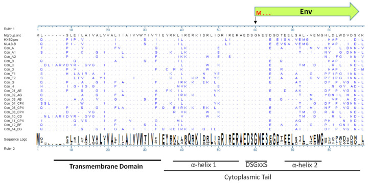
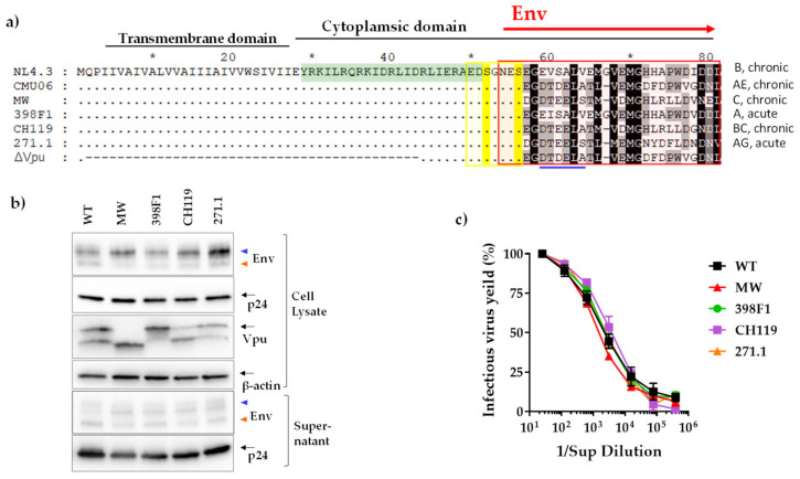
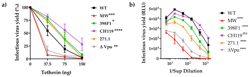
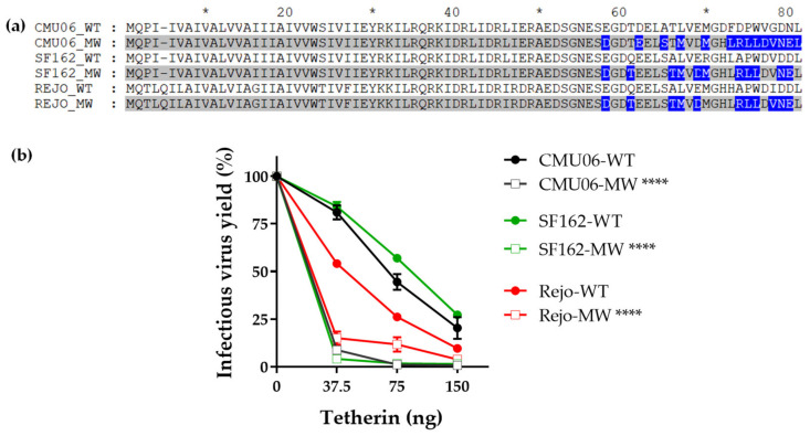
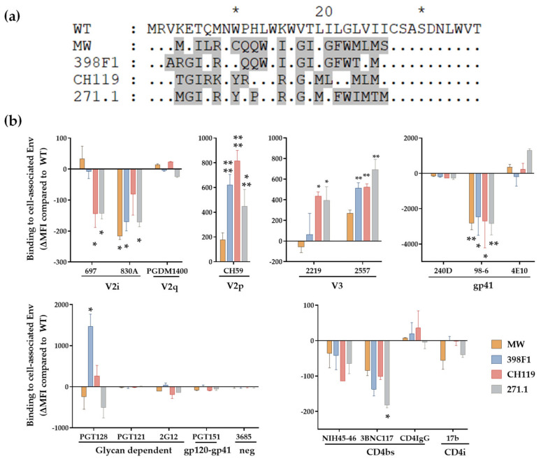
HIV-1 Env signal peptide (SP) is an important contributor to Env functions. Env is generated from Vpu/Env encoded bicistronic mRNA such that the 5' end of Env-N-terminus, that encodes for Env-SP overlaps with 3' end of Vpu. Env SP displays high sequence diversity, which translates into high variability in Vpu sequence. This study aimed to understand the effect of sequence polymorphism in the Vpu-Env overlapping region (VEOR) on the functions of two vital viral proteins: Vpu and Env. We used infectious molecular clone pNL4.3-CMU06 and swapped its SP (or VEOR) with that from other HIV-1 isolates. Swapping VEOR did not affect virus production in the absence of tetherin however, presence of tetherin significantly altered the release of virus progeny. VEOR also altered Vpu's ability to downregulate CD4 and tetherin. We next tested the effect of these swaps on Env functions. Analyzing the binding of monoclonal antibodies to membrane embedded Env revealed changes in the antigenic landscape of swapped Envs. These swaps affected the oligosaccharide composition of Env-N-glycans as shown by changes in DC-SIGN-mediated virus transmission. Our study suggests that genetic diversity in VEOR plays an important role in the differential pathogenesis and also assist in immune evasion by altering Env epitope exposure.

Keywords: DC-SIGN; HIV-1; Vpu; antibodies; envelope; glycosylation; tetherin antagonism; transmission.


Conflict of interest statement

The authors declare no conflict of interest.

Figures

Figure 1
Figure 1
Alignment of consensus sequence of Vpu from HIV-1 group M subtypes showing amino acid variability especially in the C-terminus. Source: Los Alamos HIV Database at https://www.hiv.lanl.gov/content/sequence/HIV/mainpage.html (accessed on 1 January 2022). The first amino acid in Env open reading frame methionine is denoted by M.
Figure 2
Figure 2
Effect of Vpu C-term swaps. (a) Amino acid alignment with reference to NL4.3. Similarity is shown by dot, insertion/deletion by trace, *, denotes blocks of 10. Important functional domains are indicated above the sequences. The canonical sorting motif (ExxxLV) is shown by blue line and the di-serine motif is highlighted in yellow. The region designated as C-terminus (VEOR) in this study is enclosed in red box. ΔVpu is CMU06 with deleted Vpu region shown in traces, similarity is shown in dots. Vpu region highlighted in green was used to immunize rabbit for producing polyclonal Ab. Infectious viruses were produced by transfecting 293T cells, that lack tetherin, with infectious molecular clones (IMC) of CMU06 proviral construct harboring chimeric Vpus and were assessed for (b) protein expression and (c) virus titer. The anti-gp120 MAb cocktail detected two Env bands corresponding to gp120 and gp160 shown by orange and blue triangle, respectively. β-actin was used as loading controls for cell lysates.
Figure 3
Figure 3
Effect of Vpu swaps on virus release in presence of tetherin. (a) Virus release from 293T cells following co-transfection with IMCs in presence of various amounts of plasmid expressing human tetherin. Release of infectious virus was determined by infection of TZM-bl indicator cells and is shown as percentage of the release efficiency in the absence of tetherin, set at 100%. Infections were performed in triplicate and mean and SEM from three experiments are shown *, p = 0.03; **, p = 0.002; ***, p = 0.0002; ****, p < 0.0001 by two-way ANOVA vs. WT. (b) Virus release from TZM.bl cells, expressing endogenous tetherin, following transfection with proviral IMC bearing the chimeric Vpu and Env. Virus release was assessed by infecting TZM.bl cells with the supernatant from each transfection. Mean +SEM from representative experiment performed in duplicate are shown. ****, p < 0.0001 by two-way ANOVA.
Figure 4
Figure 4
Down modulation of tetherin and CD4. (a) TZM.bl cells expressing endogenous tetherin and CD4 were transfected with CMU06 proviral plasmids expressing chimeric Vpu and Env. Cells were analyzed for tetherin and CD4 expression 48h post-transfection by flow cytometry. Median fluorescent intensity (MFI) + SD values of triplicates from representative experiment are shown. *, p <0.05; **, p < 0.01; ****, p < 0.0001 by one-way ANOVA vs. WT. (b) Spearman correlation between downmodulation of surface tetherin or CD4 and virus release in TZM.bl cells (AUC from infectivity curves in Figure 3).
Figure 5
Figure 5
Effect of Vpu C-term swap on virus release in presence of tetherin. Vpu C-term MW965.26 was swapped in context of different isolates including CMU06 (acute), SF162 (chronic) and Rejo (T/F). (a) Alignment showing the differences in sequences. Sequences with swapped Vpu C-terminus are shaded in gray and the difference between the MW and WT sequences are highlighted in blue, gaps are indicated by trace. *, denotes blocks of 10. (b) HEK293T cells were co-transfected with IMCs and various amounts of plasmid expressing human tetherin. Forty-eight hours post-transfection supernatants were tested for infectious virus release by infecting TZM.bl cells as in Figure 2. ****, p < 0.0001 by ANOVA compared to their respective WT.
Figure 6
Figure 6
Effect of SP swap on CMU06 Env-antibody interaction. (a) Alignment showing the differences in SP sequences used to express CMU06 Env. SP sequences different from CMU06 native WT SP are shaded in gray and the similarities between the SP are indicated by dot, * denote blocks of 10. (b) Binding of mAbs to WT vs. SP-swapped Envs expressed on the surface of HEK293T cells. The mAbs were tested at following concentrations: 697, 830, and 3685 at 100 μg/mL; 2G12 at 50 μg/mL; PGDM1400, 2219, 2557, 240D, 98-6 and PGT121 at 25 μg/mL; 4E10, 17b at 10 μg/mL; PGT128, PGT151, 3BNC117 and CD4-IgG at 5 μg/mL; CH59 and NIH45-46 at 2.5 μg/mL. Normalized geometric mean fluorescence intensity (MFI) compared to WT (set as 100%) and SD from duplicates in one experiment are shown. MFI of negative control mAb were subtracted. Data were analyzed by ANOVA (*, p < 0.05; **, p < 0.01; *** p < 0.001; ****, p < 0.0001 vs. WT).
Figure 7
Figure 7
Effect of SP swap on REJO and SF162 Env-antibody interaction. HEK293T cells transfected with REJO (a) and SF162 (b) gp160 were analyzed for binding to mAbs targeting different Env epitopes. The mAbs were tested at following concentrations: 697, 830, and 3685 at 100 μg/mL; 2G12 at 50 μg/mL; PGDM1400, 2219, 2557, 240D, 98-6 and PGT121 at 25 μg/mL; 4E10, 17b at 10 μg/mL; PGT128, PGT151, 3BNC117 and CD4-IgG at 5 μg/mL; CH59 and NIH45-46 at 2.5 μg/mL. Normalized geometric mean fluorescence intensity (MFI) compared to WT (set as 100%) and SD from duplicates in one experiment are shown. MFI of negative control mAb were subtracted. Data were analyzed by ANOVA (*, p < 0.05; **, p < 0.01; *** p < 0.001; vs. WT). Sequence differences among the respective WT and swapped SP are also shown. Similar residues are shown as dot, * denote blocks of 10.
Figure 8
Figure 8
DC-SIGN mediated HIV-1 transmission. Raji–DC-SIGN+ cells were incubated for 2 h with WT or SP swapped viruses produced in (a) HEK293T and (b) human peripheral blood mononuclear cells (PBMC). Cells were washed to remove unbound viruses and added to CD4+ TZM.bl cells. Viral transmission to the TZM-bl cells was determined by luciferase activity and calculated based on infection in TZM.bl cells without Raji cells as control (set to 100%). Background luciferase activity was determined in co-cultures without any virus. *, p < 0.05; **, p < 0.01; ***, p < 0.001; ****, p < 0.0001 vs. by ANOVA.

Similar articles

Cited by

References

    1. Cohen E.A., Terwilliger E.F., Sodroski J.G., Haseltine W.A. Identification of a protein encoded by the vpu gene of HIV-1. Nature. 1988;334:532–534. doi: 10.1038/334532a0. - DOI - PubMed
    1. Kmiec D., Iyer S.S., Stürzel C.M., Sauter D., Hahn B., Kirchhoff F. Vpu-Mediated Counteraction of Tetherin Is a Major Determinant of HIV-1 Interferon Resistance. mBio. 2016;7:e00934-16. doi: 10.1128/mBio.00934-16. - DOI - PMC - PubMed
    1. Schwartz S., Felber B.K., Fenyö E.M., Pavlakis G.N. Env and Vpu proteins of human immunodeficiency virus type 1 are produced from multiple bicistronic mRNAs. J. Virol. 1990;64:5448–5456. doi: 10.1128/jvi.64.11.5448-5456.1990. - DOI - PMC - PubMed
    1. Upadhyay C., Feyznezhad R., Yang W., Zhang H., Zolla-Pazner S., Hioe C.E. Alterations of HIV-1 envelope phenotype and antibody-mediated neutralization by signal peptide mutations. PLoS Pathog. 2018;14:e1006812. doi: 10.1371/journal.ppat.1006812. - DOI - PMC - PubMed
    1. Vermeire J., Roesch F., Sauter D., Rua R., Hotter D., Van Nuffel A., Vanderstraeten H., Naessens E., Iannucci V., Landi A., et al. HIV Triggers a cGAS-Dependent, Vpu- and Vpr-Regulated Type I Interferon Response in CD4+ T Cells. Cell Rep. 2016;17:413–424. doi: 10.1016/j.celrep.2016.09.023. - DOI - PubMed

Publication types

MeSH terms

Substances