. 2022 Apr 10;27(8):2438.
doi: 10.3390/molecules27082438.

Reactive Acrylamide-Modified DNA Traps for Accurate Cross-Linking with Cysteine Residues in DNA-Protein Complexes Using Mismatch Repair Protein MutS as a Model

Affiliations

Reactive Acrylamide-Modified DNA Traps for Accurate Cross-Linking with Cysteine Residues in DNA-Protein Complexes Using Mismatch Repair Protein MutS as a Model

Mayya V Monakhova et al. Molecules. .

Abstract

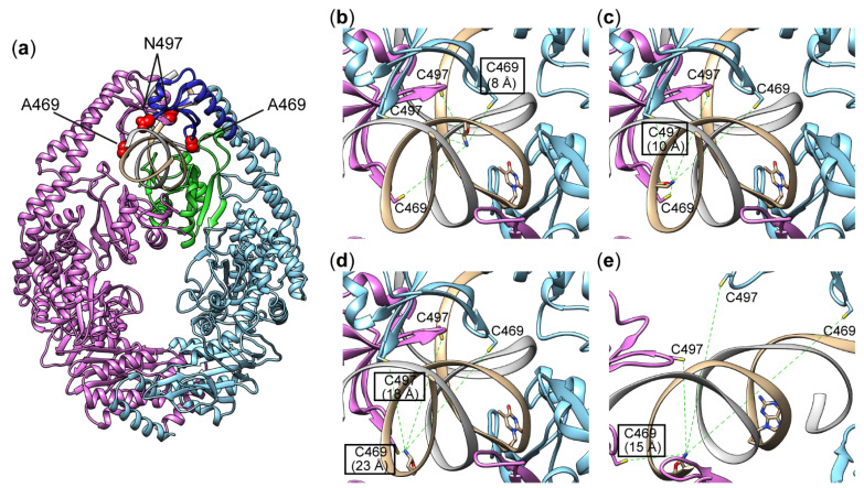
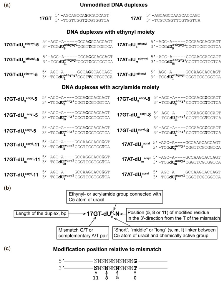
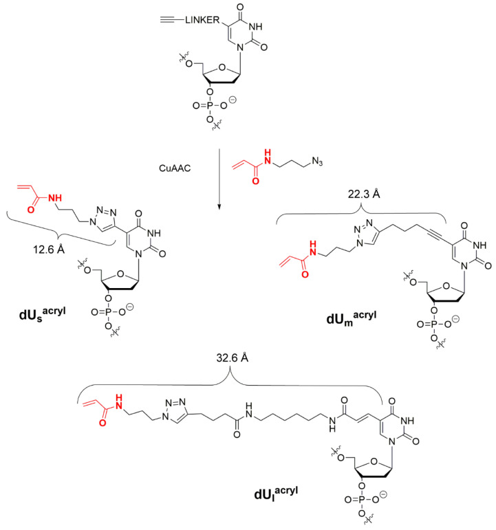
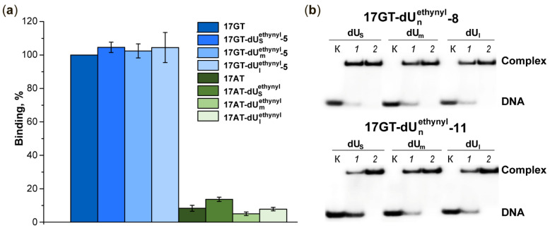
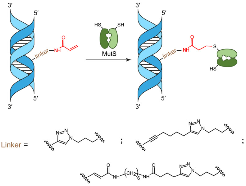
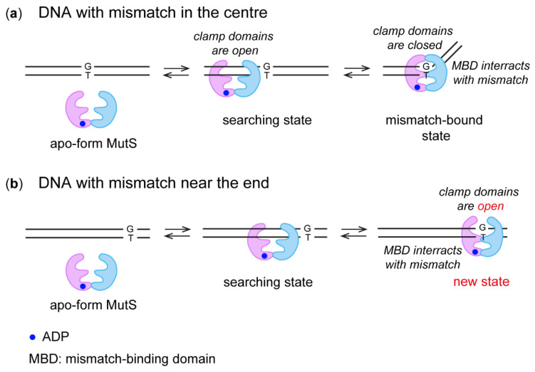
Covalent protein capture (cross-linking) by reactive DNA derivatives makes it possible to investigate structural features by fixing complexes at different stages of DNA-protein recognition. The most common cross-linking methods are based on reactive groups that interact with native or engineered cysteine residues. Nonetheless, high reactivity of most of such groups leads to preferential fixation of early-stage complexes or even non-selective cross-linking. We synthesised a set of DNA reagents carrying an acrylamide group attached to the C5 atom of a 2'-deoxyuridine moiety via various linkers and studied cross-linking with MutS as a model protein. MutS scans DNA for mismatches and damaged nucleobases and can form multiple non-specific complexes with DNA that may cause non-selective cross-linking. By varying the length of the linker between DNA and the acrylamide group and by changing the distance between the reactive nucleotide and a mismatch in the duplex, we showed that cross-linking occurs only if the distance between the acrylamide group and cysteine is optimal within the DNA-protein complex. Thus, acrylamide-modified DNA duplexes are excellent tools for studying DNA-protein interactions because of high selectivity of cysteine trapping.

Keywords: DNA mismatch repair; DNA modification; DNA–protein complex; MutS; crosslinking; modified oligonucleotide; regioselectivity.


Conflict of interest statement

The authors declare no conflict of interest.

Figures

Figure 1
Figure 1
Complexes of MutS with DNA. An overview of the structure of MutS bound to DNA containing a mismatch (a). The clamp domain is highlighted in blue; the mismatch-binding domain is green, and A469 and N497 are red. The distances from the SG atom of C469 and C497 to the N atom of the heterocyclic base in the nucleotide located at the 5th (b), 8th (c) or 11th (d) position relative to the mismatch (see Figure 2). Panels (bd) represent a detailed view of the structure of the MutS complex with a G/T-containing duplex, as determined by Cryo-EM (PDB ID 7AI6). Panel (e) is a fragment of a MutS complex with a canonical duplex (PDB ID 7AI5). The position of the modified base in the complex without the mismatch was determined by superposition of Cryo-EM structures with PDB IDs 7AI6 and 7AI5 (see Section 3.2 and Supplementary Materials). The two subunits of the protein are highlighted in blue (subunit A) and pink (subunit B), and the DNA is brown. The clamp domain is blue; the mismatch-binding domain is green, and A469 and N497 are red. Structures of native protein 7AI6 and 7AI5 have been described previously [22].
Figure 2
Figure 2
DNA duplexes used in this study. (a) The structure of DNA duplexes; modified dU residues are shown in Figure 3. (b) Explanation of the name of a duplex. (c) Illustration of positions of modified dU with respect to T of the G/T pair (position 0).
Figure 3
Figure 3
The scheme of the synthesis of oligonucleotides carrying the acrylamide group from ethynyl-containing oligonucleotides.
Figure 4
Figure 4
Binding of DNA duplexes to MutS(A469C). (a) Binding efficiency. Formation of the MutS(A469C) complex with an unmodified G/T-containing duplex is set to 100%. The standard deviation from the mean of at least three experiments is indicated; p < 0.05. (b) Analysis of complex formation between MutS(A469C) and 32P-labelled duplexes 17GT-dUsethynyl-8, 17GT-dUmethynyl-8, 17GT-dUlethynyl-8 (top panel), 17GT-dUsethynyl-11, 17GT-dUmethynyl-11 and 17GT-dUlethynyl-11 (bottom panel) by an electrophoresis mobility shift assay (EMSA). DNA concentration was 0.5 µM. MutS(A469C) concentration was 1 µM (a, lanes 1) or 2.5 µM (lanes 2). Lanes K correspond to DNA without the protein. Autoradiograph of a 6% polyacrylamide gel.
Scheme 1
Scheme 1
Affine modification of MutS with acrylamide containing DNA duplex.
Figure 5
Figure 5
Cross-linking of MutS(A469C), MutS(N497C), CFMutS or WTMutS (5 μM on the monomer basis) with DNA duplex 17GT-dUsacryl-5, 17GT-dUmacryl-5, 17GT-dUlacryl-5, 17AT-dUsacryl, 17AT-dUmacryl or 17AT-dUlacryl (0.5 μM) containing a 32P label at the 5′ end of the modified strand. Analysis by 8% polyacrylamide gel electrophoresis (PAGE) with 0.1% of SDS. Autoradiograph (a) and photographs (b,d) of the gels stained with a Coomassie G250 solution. (a,b) Lanes 16: products of MutS(A469C) cross-linking with duplex 17GT-dUsacryl-5, 17GT-dUmacryl-5, 17GT-dUlacryl-5, 17AT-dUsacryl, 17AT-dUmacryl or 17AT-dUlacryl, respectively. (c) Efficiency of a MutS(A469C) or MutS(N497C) reaction with 17GT-dUnacryl-5 or 17AT-dUnacryl. The error bars represent the standard deviation of three independent experiments; p < 0.05. (d) Interaction of CFMutS (lanes 13) or WTMutS (lanes 46) with 17GT-dUsacryl-5, 17GT-dUmacryl-5 or 17GT-dUlacryl-5. Lanes K correspond to the protein without DNA; DNA lane: 17GT, M: markers of protein molecular mass, kDa.
Figure 6
Figure 6
MutS(N469C) (a) or MutS(N497C) (b) cross-linking with 17GT-dUsacryl-5, 17GT-dUmacryl-5 or 17GT-dUlacryl-5 (lanes 13); 17GT-dUsacryl-8, 17GT-dUmacryl-8 or 17GT-dUlacryl-8 (lanes 46); or 17GT-dUsacryl-11, 17GT-dUmacryl-11 or 17GT-dUlacryl-11 (lanes 79). Conjugate yields (%) are indicated under the gel lanes. Protein concentration on the monomer basis was 5 µM, DNA: 0.5 µM. Duplexes contain a 32P label at the 5′ end of the modified strand. Autoradiograph of SDS-PAGE in an 8% gel.
Figure 7
Figure 7
Analysis of MutS(N497C) (5 μM on the monomer basis) cross-linking with the 17GT-dUmacryl-8 DNA duplex (0.5 μM) by SDS-PAGE in an 8% gel. Reaction conditions: 0–120 min, 37 °C. Conjugate yields (%) are indicated under the gel lanes. The gel was stained with a Coomassie G250 solution. M: markers of protein molecular mass, kDa.
Figure 8
Figure 8
MutS binding to DNA with a mismatch in the middle (a) or near a terminus of the duplex (b).

References

    1. Lambert M., Jambon S., Depauw S., David-Cordonnier M.-H. Targeting transcription factors for cancer treatment. Molecules. 2018;23:1479. doi: 10.3390/molecules23061479. - DOI - PMC - PubMed
    1. Jiang M., Jia K., Wang L., Li W., Chen B., Liu Y., Wang H., Zhao S., He Y., Zhou C. Alterations of DNA damage repair in cancer: From mechanisms to applications. Ann. Transl. Med. 2020;8:1685. doi: 10.21037/atm-20-2920. - DOI - PMC - PubMed
    1. Ilina E.S., Khodyreva S.N., Lavrik O.I. Unusual interaction of human apurinic/apyrimidinic endonuclease 1 (APE1) with abasic sites via the Schiff-base-dependent mechanism. Biochimie. 2018;150:88–99. doi: 10.1016/j.biochi.2018.04.027. - DOI - PubMed
    1. Khodyreva S., Lavrik O. Non-canonical interaction of DNA repair proteins with intact and cleaved AP sites. DNA Repair. 2020;90:102847. doi: 10.1016/j.dnarep.2020.102847. - DOI - PubMed
    1. Hyjek-Składanowska M., Stasińska A.R., Napiórkowska-Gromadzka A., Bartłomiejczak A., Seth P.P., Chmielewski M.K., Nowotny M. Disulfide bridge cross-linking between protein and the RNA backbone as a tool to study RNase H1. Bioorg. Med. Chem. 2020;28:115741. doi: 10.1016/j.bmc.2020.115741. - DOI - PubMed

LinkOut - more resources