. 2022 Apr 13;27(8):2515.
doi: 10.3390/molecules27082515.

Exogenous Melatonin Activating Nuclear Factor E2-Related Factor 2 (Nrf2) Pathway via Melatonin Receptor to Reduce Oxidative Stress and Apoptosis in Antler Mesenchymal Stem Cells

Affiliations

Exogenous Melatonin Activating Nuclear Factor E2-Related Factor 2 (Nrf2) Pathway via Melatonin Receptor to Reduce Oxidative Stress and Apoptosis in Antler Mesenchymal Stem Cells

Huansong Jing et al. Molecules. .

Abstract

Antler growth depends on the proliferation and differentiation of mesenchymal stem cells (MSCs), and this process may be adversely affected by oxidative stress. Melatonin (MLT) has antioxidant functions, but its role in Cervidae remains largely unknown. In this article, flow cytometry, reactive oxygen species (ROS) identification, qPCR, and other methods were used to investigate the protective mechanism of MLT in H2O2-induced oxidative stress of antler MSCs. The results showed that MLT significantly increases cell viability by relieving the oxidative stress of antler MSCs. MLT inhibits cell apoptosis by protecting mitochondrial function. We blocked the melatonin receptor with luzindole (Luz) and found that the receptor blockade significantly increases H2O2-induced hyperoxide levels and causes significant inhibition of mitochondrial function. MLT treatment activates the nuclear factor E2-related factor 2 (Nrf2) antioxidant signaling pathway, up-regulates the expression of NAD(P)H quinone oxidoreductase 1 (NQO1) and other genes and it could inhibit apoptosis. In contrast, the melatonin receptor blockade down-regulates the expression of Nrf2 pathway-related genes, but significantly up-regulates the expression of apoptotic genes. It was indicated that MLT activates the Nrf2 pathway through the melatonin receptor and alleviates H2O2-induced oxidative stress and apoptosis in antler MSCs. This study provides a theoretical basis for further studying the oxidative stress and antioxidant process of antler MSCs and, thereby, increasing antler yields.

Keywords: MSCs; Nrf2 pathway; antler; apoptosis; melatonin; oxidative stress.


Conflict of interest statement

The authors declare no conflict of interest.

Figures

Figure 1
Figure 1
Cell type identification, effects of H2O2 on cell viability, gene expression, and ROS level. (A) mRNA level of OCT3/4, CD90, and CD73 in experimental cells and EECs. (B) Cell viability determined by cck-8 method after gradient H2O2 treatment with 6 replicates per treatment. (C,D) Expression of apoptosis-related genes and antioxidant enzymes detected by qPCR with 3 replicates per treatment. (E) ROS fluorescence images captured by fluorescence-forward microscopy (n = 3, scale: 100 μm). (F) Fluorescence intensity calculated by ImageJ. H2O2 group was treated with H2O2 (400 μmol/L) for 2 h. The results were obtained from three independent experiments, and data were expressed as mean ± SD. * p < 0.05 (** p < 0.01) vs. control group; ns, not significant, p > 0.05 vs. control group.
Figure 2
Figure 2
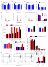
Effects of MLT treatment on cell viability, cell cycle, apoptosis, intracellular ROS, MDA level, and mitochondrial function. (A) Protective effect of gradient MLT on cell viability detected by CCK-8 method after treatment with concentrations of 400 μmol/L H2O2, n = 6. (B) Protective effect of gradient MLT on cell viability detected by CCK-8 method after treatment with concentrations of 500 μmol/L H2O2, n = 6. (C) Protective effect of gradient MLT on cell viability detected by CCK-8 method after treatment with concentrations of 600 μmol/L H2O2, n = 6. (D) Protective effect of gradient MLT on cell viability detected by CCK-8 method after treatment with concentrations of 800 μmol/L H2O2, n = 6. (E,F) Flow cytometry analysis of cell cycle in control group, H2O2 treatment group, and MLT + H2O2 group, n = 3. (G) Intracellular ROS level in gradient H2O2 group and 100 ng/mL MLT and gradient H2O2 group, n = 6. (H) MDA level under gradient H2O2 treatment and MLT + H2O2 treatment, n = 6. (I,J) Apoptosis-related gene expression and mtDNA levels in control group, H2O2 treatment group, and MLT + H2O2 group, n = 3. (K) Mitochondrial membrane potential in control group, H2O2 treatment group, and MLT + H2O2 group, n = 6. (L) Intracellular ATP level in control group, H2O2 treatment group, and MLT + H2O2 group, n = 6. (M,N) Flow cytometry analysis of cell apoptosis in control group, H2O2 treatment group, and MLT + H2O2 group, n = 3. H2O2 group, 2 h 400 μmol/L H2O2 treatment; MLT+ H2O2 group, 12 h 100 ng/mL MLT treatment, followed by MLT wash with PBS and 2 h 400 μmol/L H2O2 treatment. The results were obtained from three independent replicates. The same letters on the error bar indicate no significant difference between the groups, and different letters above error bars (a–e) represent significant differences between groups at the level of p < 0.05.
Figure 3
Figure 3
Effect of melatonin receptor blockade on alleviation of oxidative stress in antler MSCs by MLT. (A) Cell viability detected by CCK-8 method in control group, H2O2 treatment group, MLT+H2O2 group and Luz+ MLT+H2O2 group, n = 6. (B) Fluorescence intensity calculated by image-J in control group, H2O2 treatment group, MLT+H2O2 group and Luz+ MLT+H2O2 group, n = 3. (C) ROS fluorescence images captured by fluorescence forward microscopy in control group, H2O2 group, MLT+H2O2 group and Luz+ MLT+H2O2 group (scale: 200 μm). (D) Relative MDA content in control group, H2O2 group, MLT+H2O2 group and Luz+ MLT+H2O2 group. (E) Relative mtDNA expression level in control group, H2O2 group, MLT+H2O2 group and Luz+ MLT+H2O2 group, n = 3. (F) Relative MMP calculated by image-J in control group, H2O2 group, MLT+H2O2 group and Luz+ MLT+H2O2 group, n = 3. (G) MMP Fluorescence images captured by fluorescence forward microscopy in control group, H2O2 group, MLT+H2O2 group and Luz+ MLT+H2O2 group (scale: 200 μm). H2O2 group, 2 h 400 μmol/L H2O2 treatment; MLT + H2O2 group, 12 h 100 ng/mL MLT treatment, followed by MLT wash with PBS and 2 h 400 μmol/L H2O2 treatment, The results were obtained from three independent replicates. The data were expressed as mean ± SD. The same letters on the error bar mean no significant difference between groups, and different letters above error bars (a–c) represent significant differences between groups at the level of p < 0.05.
Figure 4
Figure 4
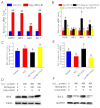
Expression of Nrf2 pathway-related and apoptosis-related genes. (A,B) Nrf2 pathway related genes expression level detected by qPCR in control group, H2O2 group, MLT+H2O2 group and Luz+ MLT+H2O2 group (n = 3). (CF) BAX, Bcl-2 expression level detected by Western blot in control group, H2O2 group, MLT+H2O2 group and Luz+ MLT+H2O2 group. H2O2 group, 2 h 400 μmol/L H2O2 treatment; MLT + H2O2 group, 12 h 100 ng/ mL MLT treatment, followed by MLT wash with PBS and 2 h 400 μmol/L H2O2 treatment; Luz + MLT + H2O2 group, 2 h 10 μmol/L luzindole treatment, followed by 12 h 100 ng/ mL MLT treatment, Luz and MLT wash with PBS, and 2 h 400 μmol/L H2O2 treatment. The results were obtained from three independent replicates. The data were expressed as mean ± SD. ** p < 0.01 vs. the control group. The same letters on the error bar mean no significant difference between the groups, and different letters above error bars (a, b) represent significant differences between groups at the level of p < 0.05.

References

    1. Li C., Suttie J.M., Clark D.E. Morphological observation of antler regeneration in red deer (Cervus elaphus) J. Morphol. 2004;262:731–740. doi: 10.1002/jmor.10273. - DOI - PubMed
    1. Goss R.J. Future directions in antler research. Anat. Rec. 1995;241:291–302. doi: 10.1002/ar.1092410302. - DOI - PubMed
    1. Kierdorf U., Kierdorf H. Deer Antlers—A Model of Mammalian Appendage Regeneration: An Extensive Review. Gerontology. 2011;57:53–65. doi: 10.1159/000300565. - DOI - PubMed
    1. Price J., Allen S. Exploring the mechanisms regulating regeneration of deer antlers. Philos. Trans. R. Soc. B Biol. Sci. 2004;359:809–822. doi: 10.1098/rstb.2004.1471. - DOI - PMC - PubMed
    1. Gyurján I., Jr., Molnár A., Borsy A., Stéger V., Hackler L., Jr., Zomborszky Z., Papp P., Duda E., Deák F., Lakatos P., et al. Gene expression dynamics in deer antler: Mesenchymal differentiation toward chondrogenesis. Mol. Genet. Genom. 2007;277:221–235. doi: 10.1007/s00438-006-0190-0. - DOI - PubMed

LinkOut - more resources