. 2022 Apr 22;22(1):114.
doi: 10.1186/s12906-022-03584-3.

Anti-SARS-CoV-2 potential of Cissampelos pareira L. identified by connectivity map-based analysis and in vitro studies

Affiliations

Anti-SARS-CoV-2 potential of Cissampelos pareira L. identified by connectivity map-based analysis and in vitro studies

Madiha Haider et al. BMC Complement Med Ther. .

Abstract

Background: Viral infections have a history of abrupt and severe eruptions through the years in the form of pandemics. And yet, definitive therapies or preventive measures are not present. Herbal medicines have been a source of various antiviral compounds such as Oseltamivir, extracted using shikimic acid from star anise (Illicium verum) and Acyclovir from Carissa edulis are FDA (Food and Drug Administration) approved antiviral drugs. In this study, we dissect the anti-coronavirus infection activity of Cissampelos pareira L (Cipa) extract using an integrative approach.

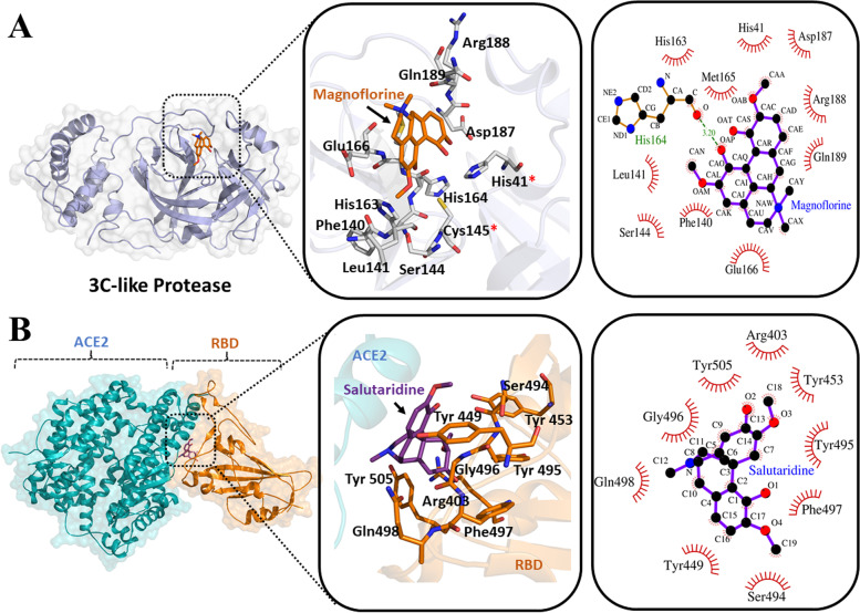
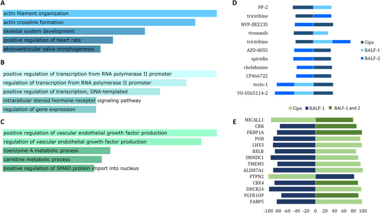
Methods: We analysed the signature similarities between predicted antiviral agents and Cipa using the connectivity map ( https://clue.io/ ). Next, we tested the anti-SARS-COV-2 activity of Cipa in vitro. Molecular docking analyses of constituents of with key targets of SARS-CoV2 protein viz. spike protein, RNA‑dependent RNA‑polymerase (RdRp) and 3C‑like proteinase. was also performed. A three-way comparative analysis of Cipa transcriptome, COVID-19 BALF transcriptome and CMAP signatures of small compounds was also performed.

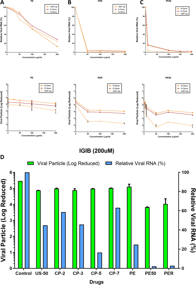
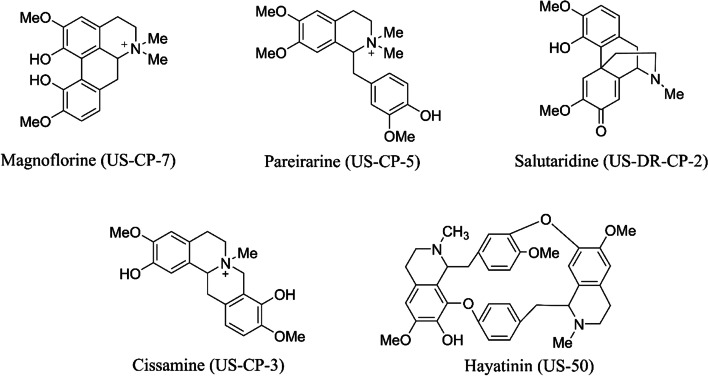
Results: Several predicted antivirals showed a high positive connectivity score with Cipa such as apcidin, emetine, homoharringtonine etc. We also observed 98% inhibition of SARS-COV-2 replication in infected Vero cell cultures with the whole extract. Some of its prominent pure constituents e.g. pareirarine, cissamine, magnoflorine exhibited 40-80% inhibition. Comparison of genes between BALF and Cipa showed an enrichment of biological processes like transcription regulation and response to lipids, to be downregulated in Cipa while being upregulated in COVID-19. CMAP also showed that Triciribine, torin-1 and VU-0365114-2 had positive connectivity with BALF 1 and 2, and negative connectivity with Cipa. Amongst all the tested compounds, Magnoflorine and Salutaridine exhibited the most potent and consistent strong in silico binding profiles with SARS-CoV2 therapeutic targets.

Keywords: Antivirus; BALF; COVID-19; Cissampelos pareira L.; Connectivity map; SARS-CoV-2; Whole plant extract.


Conflict of interest statement

The authors declare no conflict of statement.

Figures

Fig. 1
Fig. 1
Inhibition of SARS-CoV-2 in vitro by Cipa whole extract and its constituents: Relative viral RNA % and Log reduction in viral particles in vero cells upon treatment at 50, 100, 150 and 200 μg of A) whole plant aqueous extract (PE), B) root extract (PER) and C) hydro-alcoholic extracts (PE50) of Cipa. D Sars-cov-2 viral titers inhibition by Cipa constituents CP-2 Salutaridine, CP-3 Cissamine, CP-5 pareirarine, CP-7 Magnoflorine, PE aqueous extract, PE50 50% hydroalcoholic extract and PER root extract. The error bars have been represented in the figures with (p < 0.05) and the experiments were done in triplicates
Fig. 2
Fig. 2
Molecular structures of the major components of the compounds isolated from Cipa and used to determine anti-SARS-CoV-2 activity
Fig. 3
Fig. 3
Cipa principal bioactive compounds bind crucial SARS-CoV2 therapeutic targets. A represents molecular docking of Magnoflorine at the active site pocket of SARS-CoV2 3C-like protease. B represents molecular docking of Salutaridine with ACE2 interacting amino acid residues of Receptor Binding Domain (RBD) from SARS-Cov2 spike protein. The middle and the right panels of each figure represent the zoomed in view of the molecular interaction patterns of the docked ligands in three-dimensional and two-dimensional spaces respectively. In the two-dimensional interaction plots, the green dotted lines indicate hydrogen bond formation, whereas the hydrophobic interactions are represented by the spiked arcs
Fig. 4
Fig. 4
Comparative analysis of Cipa with BALF-1 and BALF-2 transcriptome: Functional enrichment of genes common between A) Cipa and BALF-1, B) Cipa and BALF-2 and C) Cipa, BALF-1 and BALF-2. D Small compound signatures common between all three, the space on the left of the vertical axis indicates negative signatures while the one on the right indicates positive signatures. E Genetic signatures common between all three, the space on the left of the vertical axis indicates negative signatures while the one on the right indicates positive signatures

Similar articles

Cited by

References

    1. Sun F, Wang X, Tan S, Dan Y, Lu Y, Zhang J, et al. SARS-CoV-2 quasispecies provides an advantage mutation pool for the epidemic variants. Microbiol Spectr. 2021;9:e0026121. doi: 10.1128/Spectrum.00261-21. - DOI - PMC - PubMed
    1. Andino R, Domingo E. Viral quasispecies. Virology. 2015;479–480:46–51. doi: 10.1016/j.virol.2015.03.022. - DOI - PMC - PubMed
    1. Planas D, Veyer D, Baidaliuk A, Staropoli I, Guivel-Benhassine F, Rajah MM, et al. Reduced sensitivity of SARS-CoV-2 variant delta to antibody neutralization. Nature. 2021;596:276–280. doi: 10.1038/s41586-021-03777-9. - DOI - PubMed
    1. Gordon DE, Jang GM, Bouhaddou M, Xu J, Obernier K, White KM, et al. A SARS-CoV-2 protein interaction map reveals targets for drug repurposing. Nature. 2020;583(7816):459–468. doi: 10.1038/s41586-020-2286-9. - DOI - PMC - PubMed
    1. Stelzig KE, Canepa-Escaro F, Schiliro M, Berdnikovs S, Prakash YS, Chiarella SE. Estrogen regulates the expression of SARS-CoV-2 receptor ACE2 in differentiated airway epithelial cells. Am J Physiol Cell Mol Physiol. 2020;318:L1280–L1281. doi: 10.1152/ajplung.00153.2020. - DOI - PMC - PubMed