Review
. 2022 Apr 22;7(1):131.
doi: 10.1038/s41392-022-00955-7.

Inflammation and atherosclerosis: signaling pathways and therapeutic intervention

Affiliations
Review

Inflammation and atherosclerosis: signaling pathways and therapeutic intervention

Peng Kong et al. Signal Transduct Target Ther. .

Abstract

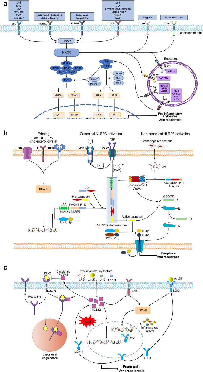
Atherosclerosis is a chronic inflammatory vascular disease driven by traditional and nontraditional risk factors. Genome-wide association combined with clonal lineage tracing and clinical trials have demonstrated that innate and adaptive immune responses can promote or quell atherosclerosis. Several signaling pathways, that are associated with the inflammatory response, have been implicated within atherosclerosis such as NLRP3 inflammasome, toll-like receptors, proprotein convertase subtilisin/kexin type 9, Notch and Wnt signaling pathways, which are of importance for atherosclerosis development and regression. Targeting inflammatory pathways, especially the NLRP3 inflammasome pathway and its regulated inflammatory cytokine interleukin-1β, could represent an attractive new route for the treatment of atherosclerotic diseases. Herein, we summarize the knowledge on cellular participants and key inflammatory signaling pathways in atherosclerosis, and discuss the preclinical studies targeting these key pathways for atherosclerosis, the clinical trials that are going to target some of these processes, and the effects of quelling inflammation and atherosclerosis in the clinic.


Conflict of interest statement

The authors declare no competing interests.

Figures

Fig. 1
Fig. 1
Key cells involved in atherosclerosis. a Phenotypic switching of VSMCs in atherosclerosis. In the healthy arterial wall, VSMCs are a contractile phenotype expressing contractile proteins (ACTA2, SM22α, Myocardin (MYOCD), and MYH11). Upon PDGF-BB and TNF-α, VSMCs switch to a synthetic phenotype, which increases the production of ECM, exosomes, pro-inflammatory cytokines, and MMPs. VSMCs release calcifying vesicles to propagate vascular calcification. KLF4 promotes phenotypic switching of VSMCs into foam-like, macrophage-like, osteochondrocyte-like, adipocyte-like, and Sca1+ mesenchymal-like VSMCs. Shear stress induces transdifferentiation of VSMCs into endothelial-like cells. The transcription factors TCF21 and OCT4 (octamer binding transcription factor) promote modulation of VSMCs into atheroprotective myofibroblast-like phenotype. b Plasticity and function of macrophages in atherosclerosis. Monocytes differentiate toward various phenotypes of macrophages in response to stimuli in atherosclerotic lesions. Among them, M1 macrophages secret pro-inflammatory cytokines; M2, M (Hb), and Mhem phenotypes are anti-inflammatory; Mox macrophages exhibit an antioxidant effect; and M4 phenotypes express pro-inflammatory cytokines and have impaired phagocytosis. c Lymphocytes in atherosclerosis. CD4+ T cells can differentiate into distinct lineages including T helper 1 (Th1), Th2, Th17, T regulatory (Treg), and many other Th cells. Th1 cells produce TNF-α and IFN-γ, indicating a pro-inflammatory and pro-atherogenic role of Th1 cells. Treg cells promote inflammatory resolution and dampen atherosclerosis progression via the production of IL-10 and TGFβ. The effect of Th2, Th9, and Th17 cells on the development of atherosclerosis remains controversial. B cells can exert both a pathogenic and protective role in atherosclerosis. B cells have two main subsets B1 and B2 cells. B1 cells exert an atheroprotective effect by the release of IgM antibodies against oxidation-specific epitopes. Similarly, Breg cells also act atheroprotective by the production of IL-10. B2 cells exhibit both pro-atherogenic and atheroprotective depending on the inflammatory microenvironment
Fig. 2
Fig. 2
Overview of inflammatory responses in atherosclerotic development. At the early stages, activated platelets mediate firm adhesion between platelets, leukocytes, and the vascular endothelium by secreting platelet-activating factor (PAF). In progressing plaques, VSMCs migrate from the medial to the subendothelial space where they undergo proliferation and developing fibrous cap. OxLDL induces phenotype switching of VSMCs to synthetic, macrophage-like VSMCs and the formation of foam cells. Excessive deposition of lipids triggers VSMC apoptosis and senescence, leading to necrosis. At predilection sites with the disturbed flow, neutrophil-released neutrophil extracellular traps (NETs) induce desquamation of endothelial cells and lead to rapid occlusion of the affected vessels. Monocyte-derived macrophages ingest oxLDL and release pro-inflammatory cytokines. Excessive deposition of lipids, as well as cytokines and histamine released from dendritic cells and mast cells, trigger the proliferation, polarization of macrophages, or even cell death
Fig. 3
Fig. 3
Key signaling pathways in atherosclerosis. a TLR signaling pathway. TLR stimulation triggers MyD88 to interact with IRAK4 (interleukin-1 receptor-associated kinase 4), which transmits signals into NF-κB and MAPK to activate the expression of inflammatory cytokines via the MyD88-dependent pathway. Endosomal TLRs transmit signals through the TRIF-dependent pathway. TRIF together with RIP1, TRAF6, and Pellino-1 activate TAK1 or with noncanonical TBK1 (Tank-binding-kinase 1) and IKKε (IKKi) activates interferon regulatory factor 3 (IRF3) and 7 to induce inflammatory cytokines. b NLRP3 inflammasome pathways. The activation of NLRP3 inflammasome has two steps: the priming and the activation. The priming is triggered by endogenous cytokines or microbial molecules. The activation of NLRP3 inflammasome includes canonical and noncanonical pathways. The canonical activation induces caspase-1 activation, which processes pro-IL-1β/pro-IL-18 to IL-1β/IL-18 active form. The noncanonical activation induced by mouse caspase-11, and human caspase-4 and caspase-5, indirectly promotes the expression of pro-IL-1β/pro-IL-18. Activated caspase-1 and caspase-11 can cleave GSDMD (gasdermin D), leading to the formation of pores in the plasma membrane and causing pyroptosis and the release of IL-1β/IL-18. c PCSK9 pathways. The expression of PCSK9 can be activated by oxLDL, LPS, and pro-inflammatory cytokines in ECs, VSMCs, and macrophages. In the absence of PCSK9, the LDL–LDLR complex is internalized. Subsequently, internalized LDLR-LDL-C complex dissociates and LDLR is recycled to the cell surface, whereas LDL-C is directed to lysosomes for degradation. PCSK9 can mediate internalized LDLR-LDL-C complex to degrade, and promote oxLDL-induced inflammation through increasing expression of LOX-1 and TLR4, which increases oxLDL uptake and upregulates inflammatory cytokine expression via activation of ROS and NF-κB

References

    1. Basatemur, G. L. et al. Vascular smooth muscle cells in atherosclerosis. Nat. Rev. Cardiol.16, 727–744 (2019). - PubMed
    1. Soehnlein O, Libby P. Targeting inflammation in atherosclerosis - from experimental insights to the clinic. Nat. Rev. Drug Discov. 2021;20:589–610. doi: 10.1038/s41573-021-00198-1. - DOI - PMC - PubMed
    1. Roy, P., Orecchioni, M. & Ley, K. How the immune system shapes atherosclerosis: roles of innate and adaptive immunity. Nat. Rev. Immunol. (2021). [Online ahead of print] - PMC - PubMed
    1. Hansson GK, Hermansson A. The immune system in atherosclerosis. Nat. Immunol. 2011;12:204–212. doi: 10.1038/ni.2001. - DOI - PubMed
    1. Ilatovskaya DV, Halade GV, DeLeon-Pennell KY. Adaptive immunity-driven inflammation and cardiovascular disease. Am. J. Physiol. Heart Circ. Physiol. 2019;317:H1254–H1257. doi: 10.1152/ajpheart.00642.2019. - DOI - PMC - PubMed

Publication types

MeSH terms

Substances