. 2022 Apr 22;10(1):65.
doi: 10.1186/s40168-022-01260-9.

Human endogenous retrovirus K in the respiratory tract is associated with COVID-19 physiopathology

Affiliations

Human endogenous retrovirus K in the respiratory tract is associated with COVID-19 physiopathology

Jairo R Temerozo et al. Microbiome. .

Abstract

Background: Critically ill 2019 coronavirus disease (COVID-19) patients under invasive mechanical ventilation (IMV) are 10 to 40 times more likely to die than the general population. Although progression from mild to severe COVID-19 has been associated with hypoxia, uncontrolled inflammation, and coagulopathy, the mechanisms involved in the progression to severity are poorly understood.

Methods: The virome of tracheal aspirates (TA) from 25 COVID-19 patients under IMV was assessed through unbiased RNA sequencing (RNA-seq), and correlation analyses were conducted using available clinical data. Unbiased sequences from nasopharyngeal swabs (NS) from mild cases and TA from non-COVID patients were included in our study for further comparisons.

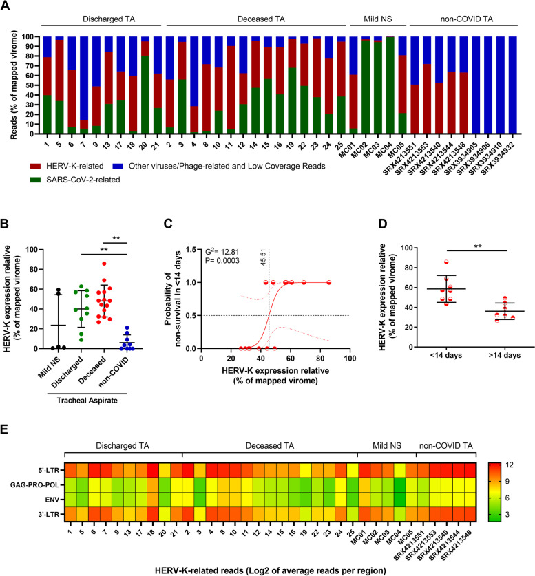
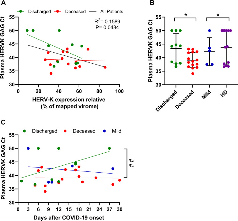
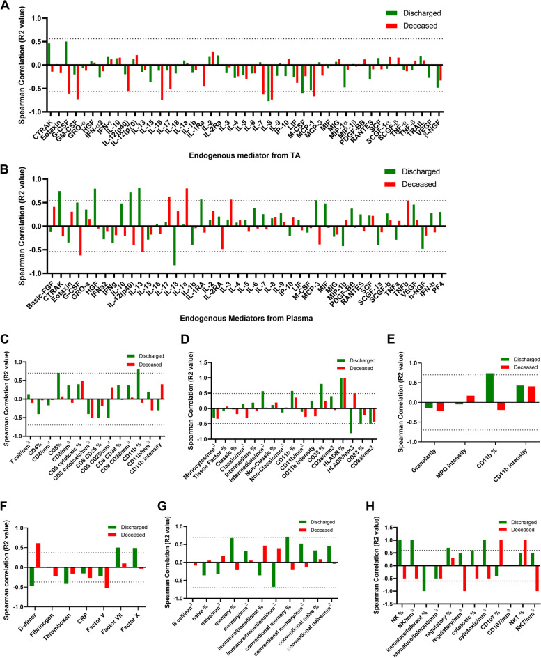
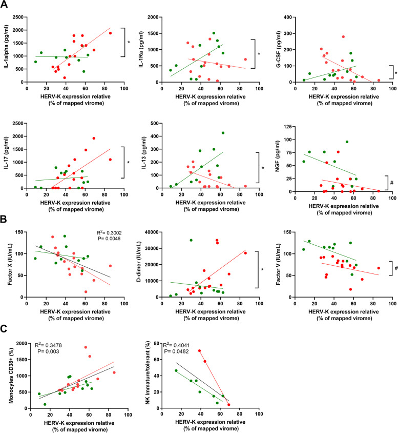
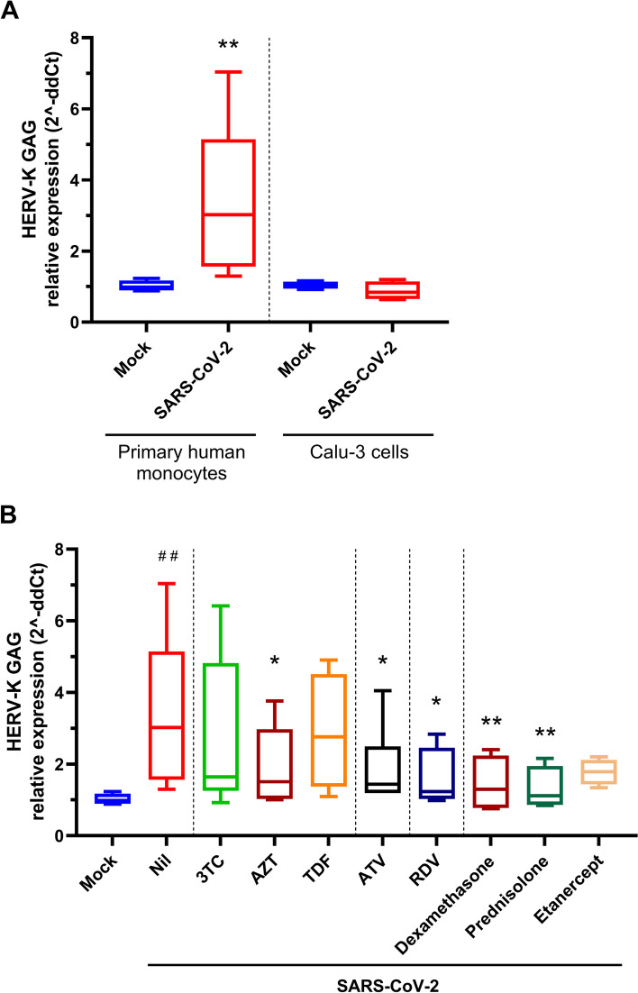
Results: We found higher levels and differential expression of human endogenous retrovirus K (HERV-K) genes in TA from critically ill and deceased patients when comparing nasopharyngeal swabs from mild cases to TA from non-COVID patients. In critically ill patients, higher HERV-K levels were associated with early mortality (within 14 days of diagnosis) in the intensive care unit. Increased HERV-K expression in deceased patients was associated with IL-17-related inflammation, monocyte activation, and an increased consumption of clotting/fibrinolysis factors. Moreover, increased HERV-K expression was detected in human primary monocytes from healthy donors after experimental SARS-CoV-2 infection in vitro.

Conclusion: Our data implicate the levels of HERV-K transcripts in the physiopathology of COVID-19 in the respiratory tract of patients under invasive mechanical ventilation. Video abstract.


Conflict of interest statement

The authors declare that they have no competing interests.

Figures

Fig. 1
Fig. 1
Differential overexpression of HERV-K transcripts in the lower respiratory tract of critically ill COVID-19 patients is associated with early mortality. RNA sequencing of tracheal aspirates (TA) from severe cases (Supplementary Table 1) and nasopharyngeal swabs (NS) from mild cases17 was performed on the MGI-2000 RNA-seq platform, and high-quality sequences (Q ≥ 30) were selected for downstream analysis. A Percentage of virus-related reads in the mapped virome from the TA of severe COVID-19 patients, from the NS from COVID-19 mild cases, and from non-COVID TA Sequence Read Archive (SRA) (# SRX4213540, SRX4213544, SRX4213548, SRX4213551, SRX4213553, SRX3934905, SRX3934906, SRX3934910, and SRX3934932). B The percentage of HERV-K-related reads in the mapped virome from the TA of discharged and deceased severe COVID-19 patients compared to NS and non-COVID TA (# SRX4213540, SRX4213544, SRX4213548, SRX4213551, SRX4213553, SRX3934905, SRX3934906, SRX3934910, and SRX3934932). C Logistic regression analysis between HERV-K expression and odds of early (< 14 days) mortality in deceased COVID-19 patients. Red dotted lines represent the 95% CI, while black dotted lines mark the intersection where data in x-axis represent 0.5 (50%) probability. Insert receiver operating characteristic (ROC) curve for the prediction of early (< 14 days) mortality in deceased COVID-19 patients based on HERV-K expression. D HERV-K expression in TA over time (days) from ICU admission to death. E Heatmap of absolute HERV-K read counts for TA from severe COVID-19 patients, NS from mild cases and for the non-COVID TA with HERV-K presence. **= p < 0.01
Fig. 2
Fig. 2
Presence of HERV-K transcripts in the plasma of severe COVID-19 patients. A The fraction of the HERV-K virome was compared to the results of real-time RT-qPCR to detect HERV-K GAG (Ct values) in plasma from those patients. B Plasma samples from severe cases (Supplementary Table 1) and from healthy donors (HD) were evaluated for the presence of HERV-K GAG by RT-qPCR. Samples with Ct values below 50 were considered positive for HERV-K. C HERV-K levels in the plasma of patients presented as a function of days since COVID-19 onset. A statistically significant (p < 0.05) difference between linear coefficients is represented by #. *= p < 0.05
Fig. 3
Fig. 3
Spearman correlation between HERV-K and severity markers in COVID-19 patients. Endogenous mediators in the TA (A), in the plasma from peripheral blood (B), T cells (C), monocytes (D), neutrophils (E), coagulation markers in the plasma (F), B cells (G), and natural killer (NK) cells (H) from peripheral blood were plotted as a function of HERV-K expression. Spearman correlation R2 was plotted, and statistical significance with p-values < 0.05 is presented by the bars that cross the dotted lines. Gate strategy for immune cell profiling is presented in Supplementary Fig. 5
Fig. 4
Fig. 4
HERV-K levels correlate with immune activation and coagulopathy in a patient outcome-dependent manner. HERV-K levels are presented as the function of A cellular survival/differentiation factors or interleukins, B clotting or fibrinolysis cascade markers, and C immune cells. These are the statistically significant analyses from Fig. 3 (panels in A and B derived from Fig. 3 A and C, respectively; panels in C derived from Fig. 3 D and H). Patients and regression lines are highlighted in green for discharged and in red for deceased patients. Regression lines in black indicate statistical significance when combining both discharged and deceased patients. Statistically significant (p < 0.05) differences between linear or angular coefficients are represented by # or *, respectively
Fig. 5
Fig. 5
Engagement of HERV-K expression by SARS-CoV-2 infection. A Human primary monocytes or Calu-3 cells were infected with an MOI of 0.1. B Human primary monocytes or Calu-3 cells were infected with an MOI of 0.1 and treated with antivirals (10 μM each) or anti-inflammatory drugs (10 μM dexamethasone and prednisolone, 25 ng/mL etanercept). A and B At 24-h postinfection, cells were lysed, and total RNA was used to quantify HERV-K GAG and RPL19 (as a reference gene). Data are presented as relative expression following the 2^-ddCt procedure. Human primary monocytes (n = 5, 2 technical replicates), Calu-3 cells (n = 3, 2 technical replicates); *= p < 0.05; **= p < 0.01

References

    1. Dong E, Du H, Gardner L. An interactive web-based dashboard to track COVID-19 in real time. Lancet Infect Dis. 2020;20:533–534. - PMC - PubMed
    1. Benefield AE, Skrip LA, Clement A, Althouse RA, Chang S, Althouse BM. SARS-CoV-2 viral load peaks prior to symptom onset: a systematic review and individual-pooled analysis of coronavirus viral load from 66 studies. medRxiv. 2020:2020.09.28.20202028.
    1. Henry BM, Aggarwal G, Wong J, Benoit S, Vikse J, Plebani M, et al. Lactate dehydrogenase levels predict coronavirus disease 2019 (COVID-19) severity and mortality: a pooled analysis. Am J Emerg Med. 2020;38:1722–1726. - PMC - PubMed
    1. Laing AG, Lorenc A, Del Barrio IDM, Das A, Fish M, Monin L, et al. A dynamic COVID-19 immune signature includes associations with poor prognosis. Nat Med. 2020;26:1623–1635. - PubMed
    1. Dorward DA, Russell CD, Um IH, Elshani M, Armstrong SD, Penrice-Randal R, et al. Tissue-specific immunopathology in fatal COVID-19. Am J Respir Crit Care Med. 2021;203:192–201. - PMC - PubMed

Publication types