. 2022 Apr 22;21(1):102.
doi: 10.1186/s12943-022-01574-0.

Antibody-mediated delivery of a viral MHC-I epitope into the cytosol of target tumor cells repurposes virus-specific CD8+ T cells for cancer immunotherapy

Affiliations

Antibody-mediated delivery of a viral MHC-I epitope into the cytosol of target tumor cells repurposes virus-specific CD8+ T cells for cancer immunotherapy

Keunok Jung et al. Mol Cancer. .

Abstract

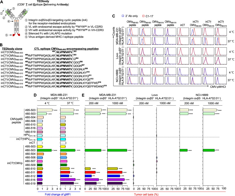
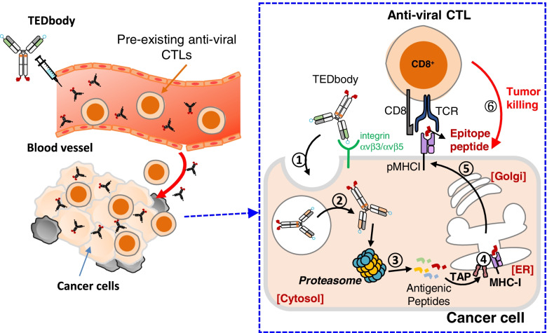
Background: Redirecting pre-existing virus-specific cytotoxic CD8+ T lymphocytes (CTLs) to tumors by simulating a viral infection of the tumor cells has great potential for cancer immunotherapy. However, this strategy is limited by lack of amenable method for viral antigen delivery into the cytosol of target tumors. Here, we addressed the limit by developing a CD8+ T cell epitope-delivering antibody, termed a TEDbody, which was engineered to deliver a viral MHC-I epitope peptide into the cytosol of target tumor cells by fusion with a tumor-specific cytosol-penetrating antibody.

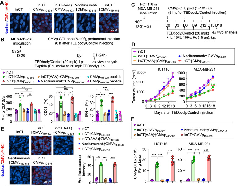
Methods: To direct human cytomegalovirus (CMV)-specific CTLs against tumors, we designed a series of TEDbodies carrying various CMV pp65 antigen-derived peptides. CMV-specific CTLs from blood of CMV-seropositive healthy donors were expanded for use in in vitro and in vivo experiments. Comprehensive cellular assays were performed to determine the presentation mechanism of TEDbody-mediated CMV peptide-MHC-I complex (CMV-pMHCI) on the surface of target tumor cells and the recognition and lysis by CMV-specific CTLs. In vivo CMV-pMHCI presentation and antitumor efficacy of TEDbody were evaluated in immunodeficient mice bearing human tumors.

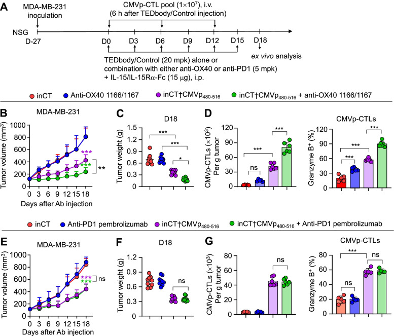
Results: TEDbody delivered the fused epitope peptides into target tumor cells to be intracellularly processed and surface displayed in the form of CMV-pMHCI, leading to disguise target tumor cells as virally infected cells for recognition and lysis by CMV-specific CTLs. When systemically injected into tumor-bearing immunodeficient mice, TEDbody efficiently marked tumor cells with CMV-pMHCI to augment the proliferation and cytotoxic property of tumor-infiltrated CMV-specific CTLs, resulting in significant inhibition of the in vivo tumor growth by redirecting adoptively transferred CMV-specific CTLs. Further, combination of TEDbody with anti-OX40 agonistic antibody substantially enhanced the in vivo antitumor activity.

Conclusion: Our study offers an effective technology for MHC-I antigen cytosolic delivery. TEDbody may thus have utility as a therapeutic cancer vaccine to redirect pre-existing anti-viral CTLs arising from previously exposed viral infections to attack tumors.

Keywords: Anti-viral cytotoxic T lymphocytes; Cytomegalovirus therapeutic cancer vaccine; Cytosol-penetrating antibody; MHC-I epitope cytosolic delivery; Peptide–MHC-I complex.


Conflict of interest statement

YSK, JAK, JK, SYL, and MJS are listed as inventors on pending patents (PCT/KR2021/001571, filed on 2 February 2021) related to the technology described in this work. All other coauthors they have no competing interests.

Figures

Fig. 1
Fig. 1
TEDbody-mediated CMV-pMHCI presentation drives efficient killing of target tumor cells by CMVp-CTLs. A Schematic of the TEDbody engineered for cytosolic delivery of an MHC-I-restricted viral CTL epitope peptide into the cytosol of target tumor cells. B Design of the TEDbodies carrying various CMVp495–503-encompassing peptides tested in this study and their nomenclature. A panel of CMVp495–503-encompassing peptides with N-terminally or N/C-extended sequences that were fused to the C-terminus of the heavy chain of inCT via an uncleavable 5-mer G4S linker. C A representative flow cytometric histogram of CMV-pMHCI display on the surface of cells as a consequence of extracellular treatment with the indicated synthetic peptide, TEDbody, or control Ab, as detected by the CMV-pMHCI-specific C1–17 Ab (red), compared to the control that used only the secondary Ab (blue). D Fold changes in the gMFI of CMV-pMHCI display, as determined by normalization to gMFI of the control involving only the secondary Ab. In (C) and (D), the indicated cells were treated with the indicated synthetic peptide, TEDbody, or control Ab (4 μM) at 4 °C for 3 h or at 37 °C for 18 h followed by flow cytometric analysis. All flow cytometric data for MDA-MB-231, LoVo, and NCI-H889 cells are shown in Fig. S3. In (D), bar graphs present the mean ± SEM (n ≥ 3). **P < 0.01 and ***P < 0.001 compared with HPVE11–19-treated cells for CMV pp65-derived peptides or compared with inCT-treated cells for the TEDbody/control Ab. E Percentage rates of tumor cell lysis by ex vivo-expanded CMVp-CTLs for integrin αvβ5+ HLA-A*02:01+ MDA-MB-231 cells, integrin αvβ5+ HLA-A*02:01 LoVo cells, and integrin αvβ5 HLA-A*02:01+ NCI-H889 cells, treated with the indicated synthetic peptide, TEDbody, or control Ab (0.2 or 1.0 μM) at 37 °C for 12 h prior to coculture with CMVp-CTLs for 18 h at an E:T ratio of 5:1. Percentage rates of lysis were determined by LDH quantification in the supernatant. The bar graphs present the mean ± SEM (n = 3)
Fig. 2
Fig. 2
TEDbody-mediated CMV-pMHCI presentation proceeds via the conventional MHC-I antigen-processing pathway. A A representative flow cytometric histogram of CMV-pMHCI display on the surface of MDA-MB-231 cells, detected by the CMV-pMHCI-specific C1–17 Ab (red) in comparison with the control involving only the secondary Ab (blue). The cells were treated with the indicated peptide, TEDbody, or control Ab (4 μM) at 4 °C for 3 h or at 37 °C for 18 h prior to flow cytometric analysis. B Percentage rates of tumor cell lysis by ex vivo-expanded CMVp-CTLs after the cancer cells were treated with the indicated peptide, TEDbody, or control Ab (20, 100, or 500 nM) for 12 h at 37 °C, prior to coculture with CMVp-CTLs for 18 h at an E:T ratio of 5:1. C IFN-γ secretion caused by the activation of CMVp-CTLs in response to CMV-pMHCI presentation on MDA-MB-231 cells after the cells were treated with the indicated peptide, TEDbody, or control Ab (0.2 μM) for 12 h at 37 °C in the absence or presence of MG132 (20 μM), ERAP1-IN-1 (20 μM), or brefeldin A (200 nM). The bar graphs show the mean ± SEM (n ≥ 3). D A representative flow cytometric histogram of CMV-pMHCI display on the surface of wild-type and TAP1 knockout MDA-MB-231 cells (red), compared to the control involving only the secondary Ab (blue). The cells were treated with the indicated TEDbody or control Ab (4 μM) at 37 °C for 18 h prior to flow cytometric analysis with the C1–17 Ab. E Representative confocal fluorescence microscopy images of MDA-MB-231 cells treated with the indicated TEDbody or control Ab (4 μM) at 37 °C for 18 h and monitoring of colocalization of CMV-pMHCI (red) with early endosome marker EEA1 (green) or a Golgi marker called 58 K Golgi (green). Nuclei were stained with Hoechst 33342 (blue). Scale bar: 20 μm. The images are representative of three independent experiments. F Percentage rates of tumor cell lysis by ex vivo-expanded CMVp-CTLs, after the cancer cells were treated with the indicated peptide, TEDbody, or control Ab (1 μM) at 37 °C for 12 h, prior to coculture with CMVp-CTLs for 18 h at the indicated E:T ratio. G and H Real-time kinetics of TEDbody-induced cell lysis of MDA-MB-231-EGFP cells by ex vivo-expanded PKH26-labeled CMVp-CTLs (G) and representative time-lapse fluorescence microscopy images (H). MDA-MB-231-EGFP cells treated with the indicated peptide, TEDbody, or control Ab (1 μM) for 12 h and then cocultivated with PKH26-labeled CMVp-CTLs at an E:T ratio of 3:1 inside the Lionheart FX automated microscopy system for the indicated periods. Lysis of MDA-MB-231-EGFP cells (green) by PKH26-labeled CMVp-CTLs (red) was registered based on a loss of the EGFP signal. In (G), the cell index refers to green fluorescence intensity from the total cancer cell area after normalization to that from the total cancer cell area at time point 0. In (H), scale bar: 20 μm. In (B), (C), (F), and (G), error bars present the mean ± SEM (n = 3). In (B), (C), (F), and (G), *P < 0.05, **P < 0.01, and ***P < 0.001 compared with the vehicle-treated control (B,C) or inCT-treated control (F,G); ns: not significant
Fig. 3
Fig. 3
TEDbody-mediated in vivo CMV-pMHCI presentation suppresses tumor growth by redirecting adoptively transferred CMVp-CTLs to tumor cells in mice. A IHC detection of CMV-pMHCI (red) on MDA-MB-231 tumor tissues excised from NSG mice bearing a preestablished MDA-MB-231 cell-derived tumor xenograft (100–120 mm3), 24 h after a single i.p. injection of the indicated TEDbody or control Ab (20 mpk). Images are representative of three independent experiments; additional images are shown in Fig. S7A. Nuclei were stained with Hoechst 33342 (blue). Scale bar: 20 μm. B Functional phenotype analysis of CMVp-CTLs from MDA-MB-231 tumor tissues, excised from NSG mice bearing a preestablished MDA-MB-231 tumor (100–120 mm3), 24 h after a single i.p. injection of the indicated TEDbody, control Ab (20 mpk), or peptide (equivalent molar amount of 20 mpk TEDbody), followed 6 h later by peritumoral injection of ex vivo-expanded CMVp-CTLs (5 × 106 cells), as described in the upper panel. Each symbol represents a value obtained from an individual mouse. The data from inCT and inCTp480–503 were pooled from two independent experiments with at least three mice per group. The bar graphs present the mean ± SEM (n ≥ 3). C The treatment scheme for assessing in vivo antitumor efficacy of the TEDbody or a control Ab in conjunction with adoptive transfer of ex vivo-expanded CMVp-CTLs in NSG mice carrying a preestablished HCT116 cell-derived subcutaneous tumor xenograft or MDA-MB-231 cell-derived orthotopic tumor xenograft (100–120 mm3). The arrows indicate each time point for the treatment or assay. D Tumor growth, measured as the average tumor volume, in response to the indicated treatment, as described in (C). Error bars: ±SEM (n = 9 to 14 per group for HCT116 tumors, n = 8 to 13 per group for MDA-MB-231 tumors). Data were pooled from two independent experiments with at least four mice per group. E and F IHC detection of CMV-pMHCI (red) on tumor tissues (E) and the number of tumor-infiltrating CMVp-CTLs per gram of a tumor (F) excised from mice on day 3 after the last treatment, as described in (C). In (E), nuclei were stained with Hoechst 33342 (blue), and images are representative of three independent experiments; additional images are shown in Fig. S7B. Scale bar: 20 μm. The right panel shows the quantification of red fluorescence intensity, obtained by ImageJ software. Error bars, ±SD of 2 fields per tumor (n = 3 per group). In (F), bar graphs present the mean ± SEM (n ≥ 3 different tumors). In (B), (D), and (F), **P < 0.01 and ***P < 0.001 denote a significant difference between the indicated groups (B and F) or a significant difference from the inCT group (B, D, and F), as determined by one-way ANOVA with the Newman–Keuls post hoc test; ns: not significant
Fig. 4
Fig. 4
Combination of TEDbody with anti-OX40 agonistic Ab augments antitumor activity. A The treatment scheme for evaluating the effect of combination of either the TEDbody or a control Ab with either the anti-OX40 agonistic 1166/1167 Ab (anti-OX40) or the anti-PD1 antagonistic Ab (pembrolizumab: anti-PD1 in the figure) in conjunction with adoptive transfer of ex vivo-expanded CMVp-CTLs in NSG mice carrying a preestablished MDA-MB-231 orthotopic tumor xenograft (100–120 mm3). The arrows indicate each time point for a treatment or assay. B and E Tumor growth, measured as the average tumor volume, in response to the indicated treatment, as described in (A). Error bars: ±SEM (n = 9 to 11 per group). Data were pooled from two independent experiments with at least with four mice per group. C, D, F and G Tumor weight (C and F) and the number of tumor-infiltrating CMVp-CTLs per gram of a tumor and percentages of granzyme B-expressing tumor-infiltrating CMVp-CTLs (D and G), determined on day 3 after the last administration of the indicated treatment, as described in (A). Each symbol represents a value for one tumor from an individual mouse; midlines indicate the mean values. In (D) and (G), error bars denote ±SEM. In (B) to (G), *P < 0.05, **P < 0.01, and ***P < 0.001 indicate a significant difference between the indicated groups or a significant difference from the inCT group (B and E), determined by one-way ANOVA with the Newman–Keuls post hoc test; ns: not significant
Fig. 5
Fig. 5
A schematic diagram of the proposed mode of action of a TEDbody in cancer immunotherapy. A TEDbody delivers a fused MHC-I-restricted viral CTL epitope peptide into the cytosol of integrin αvβ5-expressing cancer cells to be processed for surface presentation by cognate MHC-I, thereby rendering the marked cancer cells recognizable and killable by pre-existing antiviral CTLs arising from common human viral infections in cancer patients as follows: 1) binding to the tumor-associated receptor, integrin αvβ5, for cellular internalization; 2) cytosolic localization through endosomal escape; 3) proteasomal cleavage and degradation generating CTL epitope precursor peptides; 4) ER uptake by TAP and N-terminal trimming of the precursor peptides to generate the mature CTL epitope, followed by its binding to cognate MHC-I; 5) cell surface presentation of the pMHCI through the ER-to-Golgi pathway; 6) recognition and lysis of pMHCI-presenting cells by pre-existing pMHCI-specific antiviral CTLs

References

    1. Leko V, Rosenberg SA. Identifying and targeting human tumor antigens for T cell-based immunotherapy of solid tumors. Cancer Cell. 2020;38(4):454–472. doi: 10.1016/j.ccell.2020.07.013. - DOI - PMC - PubMed
    1. Saxena M, van der Burg SH, Melief CJM, Bhardwaj N. Therapeutic cancer vaccines. Nat Rev Cancer. 2021;21(6):360–378. doi: 10.1038/s41568-021-00346-0. - DOI - PubMed
    1. Lang F, Schrors B, Lower M, Tureci O, Sahin U. Identification of neoantigens for individualized therapeutic cancer vaccines. Nat Rev Drug Discov. 2022;21(4):261‒82. - PMC - PubMed
    1. Ott PA, Hu Z, Keskin DB, Shukla SA, Sun J, Bozym DJ, Zhang W, Luoma A, Giobbie-Hurder A, Peter L, et al. An immunogenic personal neoantigen vaccine for patients with melanoma. Nature. 2017;547(7662):217–221. doi: 10.1038/nature22991. - DOI - PMC - PubMed
    1. Simoni Y, Becht E, Fehlings M, Loh CY, Koo SL, Teng KWW, Yeong JPS, Nahar R, Zhang T, Kared H, et al. Bystander CD8(+) T cells are abundant and phenotypically distinct in human tumour infiltrates. Nature. 2018;557(7706):575–579. doi: 10.1038/s41586-018-0130-2. - DOI - PubMed

Publication types