. 2022 Apr 22;11(1):19.
doi: 10.1038/s41389-022-00392-9.

TCRP1 activated by mutant p53 promotes NSCLC proliferation via inhibiting FOXO3a

Affiliations

TCRP1 activated by mutant p53 promotes NSCLC proliferation via inhibiting FOXO3a

Hao Liu et al. Oncogenesis. .

Abstract

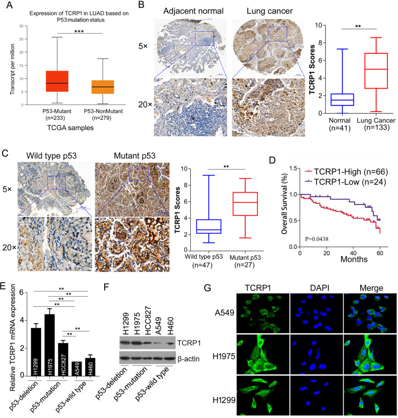
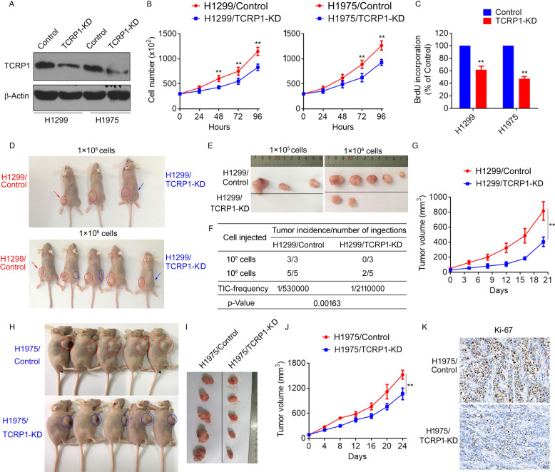
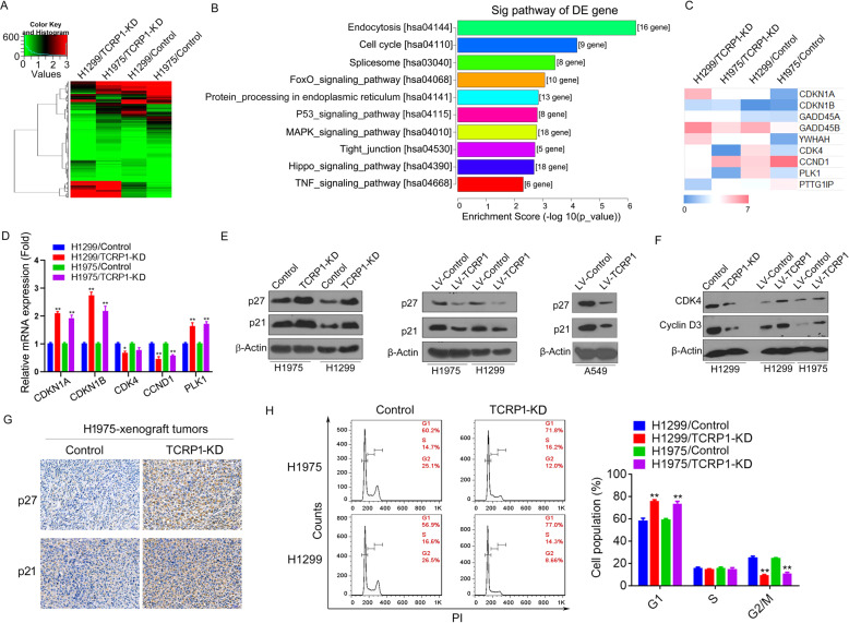
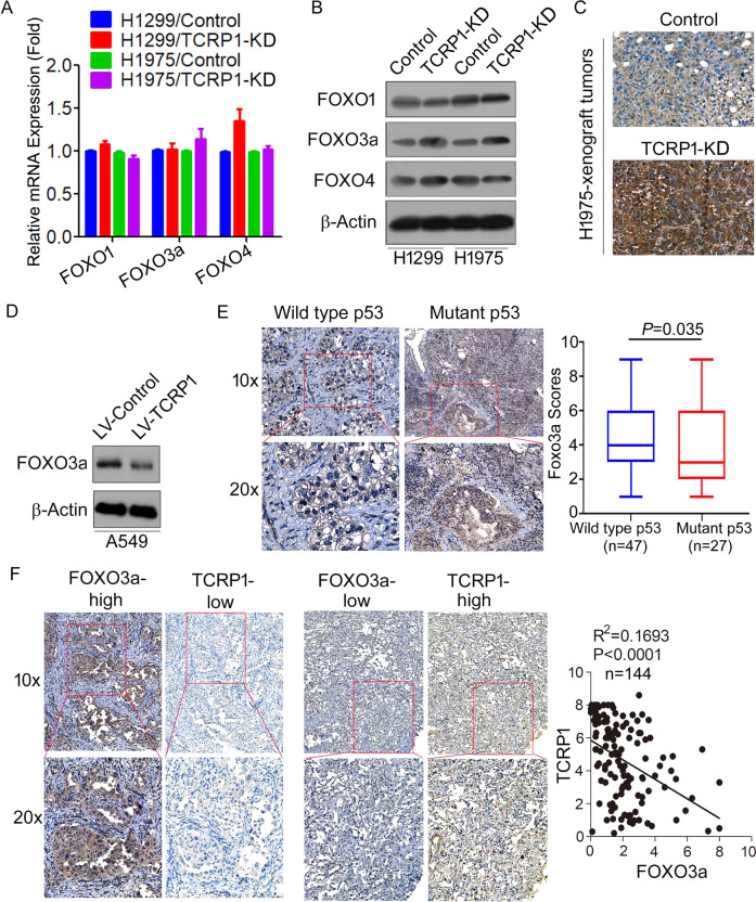
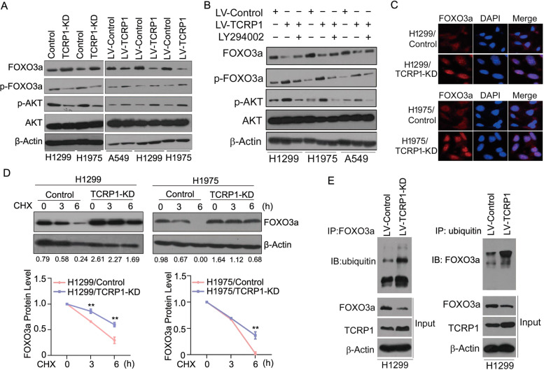
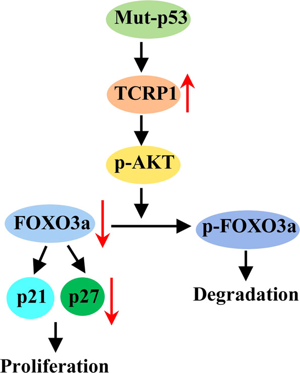
Previously, our lab explored that tongue cancer resistance-associated protein (TCRP1) plays a central role in cancer chemo-resistance and progression. Absolutely, TCRP1 was significantly increased in lung cancer. But the mechanism is far from elucidated. Here, we found that TCRP1 was increased in p53-mutant non-small-cell lung cancer (NSCLC), comparing to that in NSCLC with wild type p53. Further study showed that mutant p53 couldn't bind to the promoter of TCRP1 to inhibit its expression. While the wild type p53 did so. Next, loss-and gain-of-function assays demonstrated that TCRP1 promoted cell proliferation and tumor growth in NSCLC. Regarding the mechanism, TCRP1 encouraged AKT phosphorylation and blocked FOXO3a nuclear localization through favoring FOXO3a ubiquitination in cytoplasm, thus, promoted cell cycle progression. Conclusionly, TCRP1 was upregulated in NSCLC cells with mutant p53. TCRP1 promoted NSCLC progression via regulating cell cycle.


Conflict of interest statement

The authors declare no competing interests.

Figures

Fig. 1
Fig. 1. TCRP1 was enhanced in p53-mutant NSCLC.
A TCRP1 expression in LUAD was measured basing on p53 mutation from TCGA databanks. vs p53-mutant, ***P < 0.001. B IHC assays were performed to evaluate TCRP1 expression in 41 cases of normal tissues and 133 cases of NSCLC tissues. vs Normal, **P < 0.01. C TCRP1 expression in NSCLC tissues with wild type p53 and mutant p53 were determined via IHC assays, respectively. vs wild type p53, **P < 0.01. D The overall survival of 90 NSCLC patients with high or low expression of TCRP1 were assessed by Kaplan-Meier plot analysis. E The mRNA expression of TCRP1 were detected in lung cancer cells by qRT-PCR assays. Among these cells, H1299 is p53 deletion, H1975 and HCC827 are p53 mutation, while A549 and H460 are p53 wild type. vs A549, vs H460, **P < 0.01. F The protein expression of TCRP1 were assessed in lung cancer cells using western blot assays. G IF assays were conducted to estimate the expression of TCRP1 in NSCLC cells.
Fig. 2
Fig. 2. Mutant p53 enhanced TCRP1 in NSCLC.
A Potential binding sites of transcriptional factor p53 in the promoter of TCRP1 were predicted via online software JASPAR. B Plasmids of wild type p53 (wt-p53) and mutant p53 (p53R175H and p53V143A) were transfected into H1299 and H1975 cells, the expression of TCRP1 in mentioned cells were estimated by qRT-PCR and western blot assays. vs Con, **P < 0.01. C ShRNAs targeting p53 and control group were introduced into A549 cells, the expression of TCRP1 in these cells were estimated by qRT-PCR and western blot assays. vs sh-con, **P < 0.01. D The schematic map of p53 binding sites on the promoter of TCRP1 were exhibited. CHIP assays were carried out to illustrate whether p53 could bind to the promoter of TCRP1. E Plasmids of pGL4-wt, pGL4-mut-1, pGL4-mut-2 and pGL4-mut-all were employed in H1299 and H1975 cells combing with wild type p53 (wt-p53) or mutant p53 (p53R175H and p53V143A), respectively. In above cells, dual-luciferase reporter assays were performed to measure the luciferase activity of each group. vs pGL4-Basic, vs pGL4-wt, **P < 0.01.
Fig. 3
Fig. 3. Knocking down TCRP1 suppressed NSCLC cell proliferation in vitro and in vivo.
CRISPR-CAS9 technique was used to knock down TCRP1 in H1299 and H1975 cells. Protein expression of TCRP1 in mentioned cells was measured via western blot assay (A). The proliferation of above cells was evaluated by cell viability (B) and BrdU assays (C), vs Control, **P < 0.01. (D) A total of 1 × 105 and 1 × 106 mentioned cells were subcutaneously injected into nude mice, respectively. The H1299/Control cells were injected at left, while the H1299/TCRP1 KD cells were injected at right. The tumors obtained from these mice were exhibited (E), the tumor incidence (F) and tumor growth curve were analyzed, vs Control, **P < 0.01 (G). The H1975/Control cells were subcutaneously injected at left of nude mice, while the H1975/TCRP1 KD cells were at right. The tumors obtained from these mice were exhibited (H). The tumor growth curves were plotted using average tumor volume within each experimental group at the set time points (I). (J) IHC assays were performed to determine ki67 expression level in these tumors. vs Control, **P < 0.01. (K) Ki-67 expression in tumors of H1975/Control and H1975/TCRP1 KD group were evaluated by IHC assays.
Fig. 4
Fig. 4. TCRP1 advances cell cycles of NSCLC cells.
A Total RNAs were collected from TCRP1-knockdown H1299 and H1975 cells, as well as their control cells, respectively. RNA-sequencing assays were performed in mentioned groups. B The differently expressed (DE) genes in above groups were analyzed by GO-KEGG analysis. C The potential DE genes related to cell cycles were exhibited. D The whole RNAs from mentioned cells were obtained. The expression levels of target genes were estimated via qRT-PCR assays, respectively. vs Control, *P < 0.05, **P < 0.01. E TCRP1 was knocked down in H1299 and H1975 cells. Meanwhile, TCRP1 was overexpressed in H1299, H1975 and A549 cells. Western blot assays were performed to evaluate p21 and p27 protein expression in each group. F The protein levels of CKD4 and Cyclin D3 were determined via western blot assays in H1299 cells with increasing or decreasing TCPR1 and H1975 cells with enhancing TCRP1. G IHC assays were carried out to measure p21 and p27 levels of H1975 cells with TCRP1 knockdown and control groups in vivo. H TCRP1 was knocked down in H1299 and H1975 cells. The cell cycle of each group was detected via flow cytometry assays. vs Control, **P < 0.01.
Fig. 5
Fig. 5. TCRP1 downregulated the protein level of FOXO3a.
In H1299 cells and H1975 cells with knocking down TCRP1 and each control cells, qRT-PCR (A) and western blot assays (B) were utilized to detect the expression of FOXO transcription factors in these cells. C The levels of FOXO3a in tumors drove by H1975 cells with reducing TCRP1 and control groups were estimated by IHC assays. D The protein levels of FOXO3a in A549 cells with restoring TCRP1 and control cells were estimated by western blot assays. E IHC assays were implemented to evaluate FOXO3a expression in 74 cases of NSCLC tissues basing on wild type p53 or mutant p53. F The expression levels of TCRP1 and FOXO3a in 144 cases of NSCLC tissues were estimated via IHC assays. The expression relationship between TCRP1 and FOXO3a was analyzed by Pearson correlation analysis.
Fig. 6
Fig. 6. TCRP1 mediated nuclear location and expression of FOXO3a via AKT.
In NSCLC cells with knocking down or increasing TCRP1 (A), and in NSCLC cells with overexpression of TCRP1 combing with PI3K inhibitor LY294002 (B), western blot assays were performed to measure the phosphorylation levels and protein levels of FOXO3a and AKT. C The nuclear localization of FOXO3a was exhibited via IF assays in H1299 and H1975 cells with decreasing TCRP1. (D) TCRP1-knockdown H1299 and H1975 cells were treated with CHX for 0, 3, and 6 h, western blot assays and Image J software were used to determine FOXO3a expression. vs Control, **P < 0.01. (E) CO-IP assays were carried out to test the ubiquitination level of FOXO3a in H1299 cells with enhancing TCRP1.
Fig. 7
Fig. 7. FOXO3a reversed the effect of TCRP1 on NSCLC cell proliferation.
shRNA targeting FOXO3a was transfected into H1299 cells and H1975 cells with decreasing TCRP1, western blot (A) and qRT-PCR assays (B) were performed to assess the expression level of FOXO3a, p21 and p27. vs TCRP1-KD, vs Control, *P < 0.01. (C) Restored overexpression of FOXO3a in A549 cells with increasing TCRP1, the protein levels of FOXO3a, p21 and p27 were determined by western blot assays. Cell viability assays (D) and BrdU assays (E) were performed to verify the proliferation ability of the mentioned cells. vs TCRP1-KD, vs Control, **P < 0.01. (F) Flow cytometry assays were employed to measure cell cycle of mentioned cells. vs TCRP1-KD, vs Control, *P < 0.01.
Fig. 8
Fig. 8
A proposed model of the mechanism by which TCRP1 enhances NSCLC cell proliferation.

Similar articles

Cited by

References

    1. Sung H, Ferlay J, Siegel RL, Laversanne M, Soerjomataram I, Jemal A, et al. Global Cancer Statistics 2020: GLOBOCAN Estimates of Incidence and Mortality Worldwide for 36 Cancers in 185 Countries. CA Cancer J Clin. 2021;71:209–49. doi: 10.3322/caac.21660. - DOI - PubMed
    1. Wender R, Fontham ET, Barrera E, Jr., Colditz GA, Church TR, Ettinger DS, et al. American Cancer Society lung cancer screening guidelines. CA Cancer J Clin. 2013;63:107–17. doi: 10.3322/caac.21172. - DOI - PMC - PubMed
    1. Dammeijer F, Lievense LA, Veerman GD, Hoogsteden HC, Hegmans JP, Arends LR, et al. Efficacy of Tumor Vaccines and Cellular Immunotherapies in Non-Small-Cell Lung Cancer: A Systematic Review and Meta-Analysis. J Clin Oncol. 2016;34:3204–12. doi: 10.1200/JCO.2015.66.3955. - DOI - PubMed
    1. Tanaka F, Ishikawa S, Yanagihara K, Miyahara R, Kawano Y, Li M, et al. Expression of angiopoietins and its clinical significance in non-small cell lung cancer. Cancer Res. 2002;62:7124–9. - PubMed
    1. Lee JJ, Park S, Park H, Kim S, Lee J, Lee J, et al. Tracing Oncogene Rearrangements in the Mutational History of Lung Adenocarcinoma. Cell. 2019;177:1842–57.e1821. doi: 10.1016/j.cell.2019.05.013. - DOI - PubMed