Osteomas of temporal bone: a rare presentation
- PMID: 35459643
- PMCID: PMC9036180
- DOI: 10.1136/bcr-2021-245334
Osteomas of temporal bone: a rare presentation
Abstract
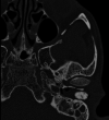
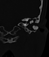
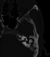
Osteoma of the temporal bone is an unusual benign slow-growing tumour composed of mature lamellar bone. It is a single pedunculated mass that often occurs unilaterally. Osteomas of external auditory canal are more common than in the other parts of temporal bone. Clinical presentation includes ear pain, hearing loss, tinnitus or vertigo. More often these lesions are an incidental finding during radiographic evaluation. Surgical excision of the osteoma is preferred in cases with impending complications. Here, we report a 36-year-old woman who came with problems of ear discharge, ear pain, hearing loss and occasional bleeding from the ear. She was diagnosed with osteoma of temporal bone with erosion of lateral semicircular canal and facial canal. Osteoma was excised and the defective areas were reconstructed.
Keywords: Ear, nose and throat; Neurootology; Pathology.
© BMJ Publishing Group Limited 2022. No commercial re-use. See rights and permissions. Published by BMJ.
Conflict of interest statement
Competing interests: None declared.
Figures


References
-
- Watkinson JC, Clarke RW, et al. Scott-Brown’s Otorhinolaryngology and Head and Neck Surgery. In: Paediatrics, the ear, and skull base surgery. Vol 2. CRC Press, 2018.
Publication types
MeSH terms
LinkOut - more resources
Full Text Sources
Medical
