

Comment
doi: 10.1136/gutjnl-2021-326617.
Epub 2022 Apr 22.
Mechanistic insight of SARS-CoV-2 infection using human hepatobiliary organoids
Affiliations
- PMID: 35459707
- PMCID: PMC9763169
- DOI: 10.1136/gutjnl-2021-326617
Item in Clipboard
Comment
Mechanistic insight of SARS-CoV-2 infection using human hepatobiliary organoids
Gut.
2023 Jan.
No abstract available
Keywords: HEPATOBILIARY DISEASE.
Conflict of interest statement
Competing interests: None declared.
Figures
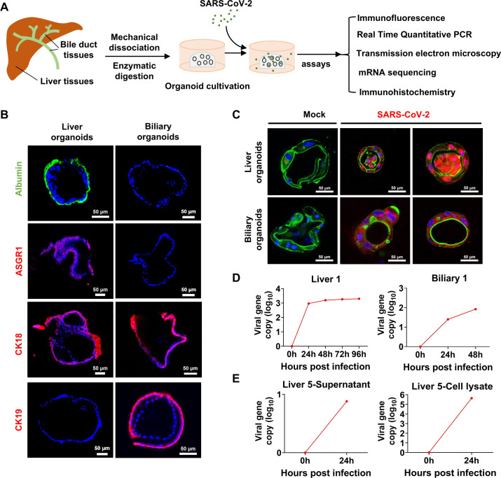
(A) Scheme of liver and biliary organoid culture establishment and performed assays. The liver and biliary organoids were generated from adjacent normal tissues of patients with liver cancer and coculture with SARS-CoV-2. (B) Differentiated liver and biliary organoids were immunostained using antibodies against human homologues to show albumin (green), ASGR1 (red), CK18 (red) and CK19 (red) protein, respectively. Nuclei are stained with DAPI (blue). Scale bar, 50 µm. (C) Immunofluorescent staining of SARS-CoV-2 infected liver organoids and biliary organoids at 24-hours. Virus are identified by SARS-CoV-2 spike (S) glycoprotein protein (red), nuclei and actin filaments are stained with DAPI (blue) and phalloidin (green), respectively. Scale bar, 50 µm. (D) Real time quantitative PCR analysis for viral sequences shows virus can productively replicate in the liver organoids at 0, 24, 48, 96 hours and in the biliary organoids at 0, 24, 48 hours after infection with SARS-CoV-2. (E) Live virus can be detected by RT-qPCR in supernatant and lysed organoids at 0 and 24 hours after infection with SARS-CoV-2.
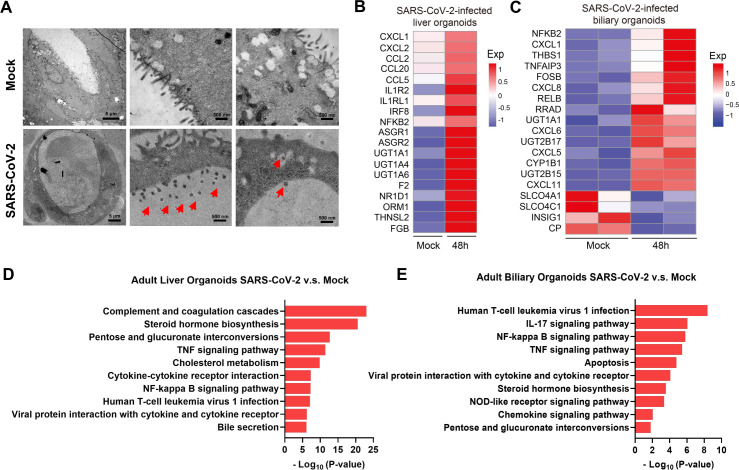
(A) Representative TEM imaging of biliary organoids at 0 and 24 hours of SARS-CoV-2 virus infection. Coronaviruses were observed in the lumen of the organoid (arrows) and are found at the membrane-bound vesicles (arrows). Scale bar 500 nm to 5 µm. (B, C) Heatmaps depicting the 19 most significantly enriched genes related to viral infection and immune system on SARS-CoV-2 infection in liver and biliary organoids. Coloured bar represents the log2-transformed values. (D, E) KEGG orthology-based annotation system of differential gene expression profiles from SARS-CoV-2-infected liver and biliary organoids compared with mock infection. TEM, transmission electron microscope.
Comment on
-
Liver function test abnormalities at hospital admission are associated with severe course of SARS-CoV-2 infection: a prospective cohort study.Gut. 2021 Oct;70(10):1925-1932. doi: 10.1136/gutjnl-2020-323800. Epub 2021 Jan 29. Gut. 2021. PMID: 33514597 Free PMC article.
References
-
- Chai X, Hu L, Zhang Y. Specific ACE2 expression in cholangiocytes may cause liver damage after 2019-nCoV infection. bioRxiv 2020.
-
- Gounden V, Vashisht R, Jialal I. Hypoalbuminemia. In: StatPearls. Treasure Island (FL): StatPearls Publishing, 2022. - PubMed
Publication types
MeSH terms
LinkOut - more resources
Full Text Sources
Medical
Miscellaneous