. 2022 May 15;208(10):2390-2402.
doi: 10.4049/jimmunol.2101085. Epub 2022 Apr 22.

Extracellular Nucleotides and Histamine Suppress TLR3- and RIG-I-Mediated Release of Antiviral IFNs from Human Airway Epithelial Cells

Affiliations

Extracellular Nucleotides and Histamine Suppress TLR3- and RIG-I-Mediated Release of Antiviral IFNs from Human Airway Epithelial Cells

Timothy S Kountz et al. J Immunol. .

Abstract

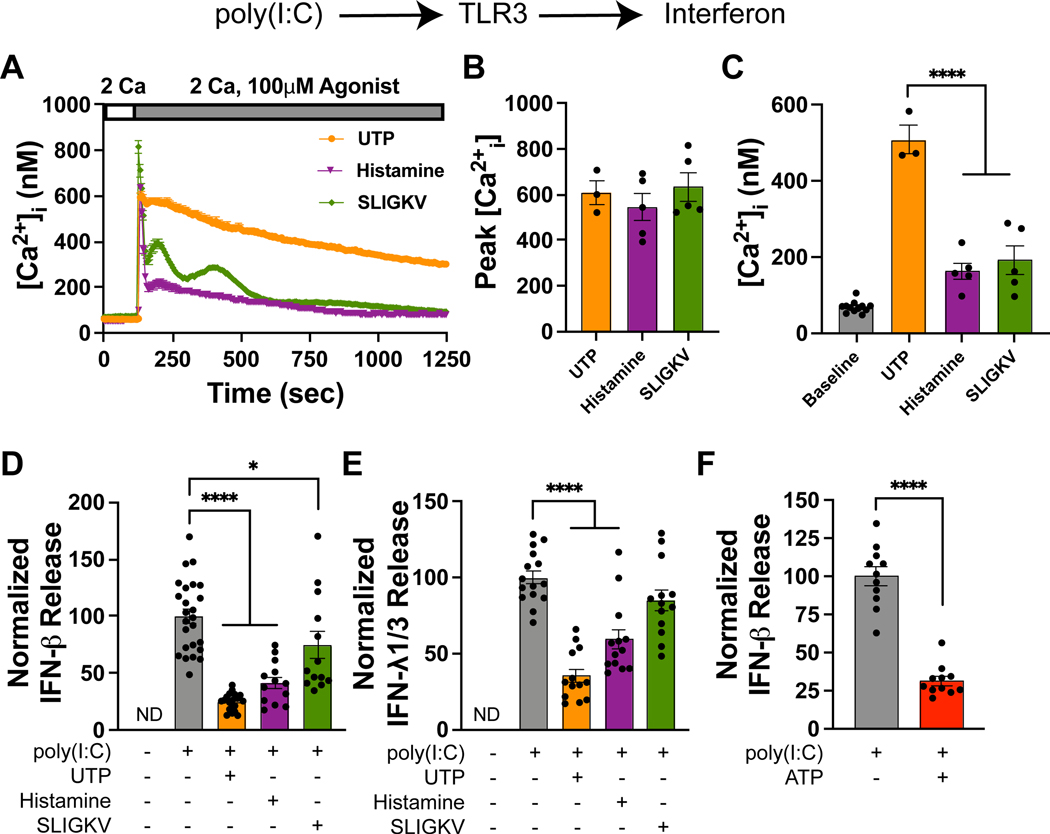
Respiratory viruses stimulate the release of antiviral IFNs from the airway epithelium. Previous studies have shown that asthmatic patients show diminished release of type I and type III IFNs from bronchial epithelia. However, the mechanism of this suppression is not understood. In this study, we report that extracellular nucleotides and histamine, which are elevated in asthmatic airways, strongly inhibit release of type I and type III IFNs from human bronchial airway epithelial cells (AECs). Specifically, ATP, UTP, and histamine all inhibited the release of type I and type III IFNs from AECs induced by activation of TLR3, retinoic acid-inducible gene I (RIG-I), or cyclic GMP-AMP synthase-STING. This inhibition was at least partly mediated by Gq signaling through purinergic P2Y2 and H1 receptors, but it did not involve store-operated calcium entry. Pharmacological blockade of protein kinase C partially reversed inhibition of IFN production. Conversely, direct activation of protein kinase C with phorbol esters strongly inhibited TLR3- and RIG-I-mediated IFN production. Inhibition of type I and type III IFNs by ATP, UTP, histamine, and the proteinase-activated receptor 2 (PAR2) receptor agonist SLIGKV also occurred in differentiated AECs grown at an air-liquid interface, indicating that the suppression is conserved following mucociliary differentiation. Importantly, histamine and, more strikingly, ATP inhibited type I IFN release from human airway cells infected with live influenza A virus or rhinovirus 1B. These results reveal an important role for extracellular nucleotides and histamine in attenuating the induction of type I and III IFNs from AECs and help explain the molecular basis of the suppression of IFN responses in asthmatic patients.


Conflict of interest statement

Competing interests

N/A.

Figures

Figure 1.
Figure 1.. UTP and histamine inhibit TLR3-induced IFN release
(A) Administration of UTP, histamine, and the PAR2 activator SLIGKV elicit [Ca2+]i elevations. [Ca2+]i was measured using Fura-2 AM as previously described (42, 96). Data are mean ± SEM of n = 40–50 cells. (B) Summary of the peak [Ca2+]i following agonist addition. Each data point is the mean peak Ca2+ signal (averaged over approximately 30–50 cells) for a given experiment (one dish) and the bar graph is mean ± SEM of n = 3–5 independent experiments. (C) Quantification of [Ca2+]i measured 5 minutes after agonist addition, reflecting Orai1-mediated SOCE. Each data point is the mean [Ca2+]i (averaged over 30–50 cells) for a given experiment (one dish) and the bar graph is the mean ± SEM of n = 3–5 independent experiments. (D) UTP (100μM), histamine (100 μM), and to a lesser degree SLIGKV inhibit poly(I:C)-induced (10μg/mL) IFN-β release into the supernatant (24-hour time point). Data are mean ± SEM of n = 13–25 samples. (E) Similarly, UTP (100 μM) and histamine (100 μM) inhibit poly(I:C)-induced (10μg/mL) IFN-λ1/3 release (24-hour time point). Data are mean ± SEM of n = 13–15 samples. (F) ATP (10μM) inhibits poly(I:C)-induced (10μg/mL) IFN-β release into the supernatant (6-hour time point). Data are mean ± SEM of n = 11 samples. This data also appears in Supplemental Fig. 1H, 3A. (A-F) All experiments were performed in primary human airway epithelial cells (NHBEs) growing in submerged cultures. *p<0.05, ****p<0.0001
Figure 2.
Figure 2.. ATP, UTP, and histamine inhibit dsRNA-induced interferon induction following mucociliary differentiation
(A) NHBEs were differentiated at the air-liquid interface (ALI) for 4 weeks and stimulated with 10μg/mL poly(I:C) and with or without the GPCR agonists (UTP, ATP, histamine, or PAR2 peptide, 100 μM each) on both apical and basolateral sides. (B and C) ALI cultures were stimulated with poly(I:C), and apical and basolateral supernatant samples were collected at the indicated time points and assessed for IFN-β (B) and IFN-λ1/3 release (C) via ELISA. Because the volumes of the apical and basolateral compartments are significantly different (~3.2-fold), the basolateral concentration was normalized to the apical side by multiplying values by 3.2. Data are mean ± SEM of n = 4–5 samples. (D-G) UTP, ATP, and histamine inhibit IFN release from dsRNA-stimulated ALI cultures. ALI cultures were stimulated with simultaneously with 10μg/mL poly(I:C) and the indicated agonists (100 μM each) on both apical and basolateral sides. The apical and basolateral supernatant samples were collected 20 hours later and measured for IFN-β (D-E) or IFN-λ1/3 (F-G) release via ELISA. Basolateral cytokine levels are shown in (D,F) while basolateral levels are shown in (E,G). Data are mean ± SEM of n = 11–21 samples. (H) ATP and UTP inhibit the release of IFN-β to both apical and basolateral compartments whereas histamine and SLIGKV exclusively inhibit basolateral IFN-β. In contrast, all agonists inhibit basolateral release of IFN-λ1/3 but not apical IFN-λ1/3 release. *p<0.05, **p<0.01, ***p<0.001, ****p<0.0001
Figure 3.
Figure 3.. P2Y2 and H1 receptors inhibit TLR3-mediated interferon release
(A) Dose-response of the inhibition of IFN-β release by UTP and histamine. IFN-β was measured in the cell culture supernatant 20 hours following simultaneous addition of poly(I:C) (10μg/mL) and either UTP or histamine. The solid line is a four-parameter nonlinear regression fit of the Hill equation with IC50 = 0.34 μM and Hill Slope = −0.52 for UTP and IC50 = 3.4 μM and Hill Slope = −1.65 for histamine. IFN-β was undetectable without poly(I:C) and hence this value (0 pg/ml) was set to 0% and the maximal poly(I:C)-evoked response was set to 100% for the fitting procedure. Data are mean ± SEM of n = 4–18 samples from 2 independent experiments. (B) Dose-response of the inhibition of IFN-λ1/3 release by UTP and histamine. IFN-λ1/3 was measured in the cell culture supernatant 20 hours following stimulation as described in (A). The solid line is a four-parameter nonlinear regression fit of the Hill equation with IC50 = 1 μM and Hill Slope = −0.7 for UTP and IC50 = 6 μM and Hill Slope = −4.09 for histamine. IFN-λ1/3 was undetectable without poly(I:C) and this value (0 pg/ml) was set to 0% and maximal poly(I:C)-evoked response was set at 100% for the fitting procedure. Data are mean ± SEM of n = 4–18 samples from 2 independent experiments. (C) The P2Y2 antagonist, AR-C 118925XX (10μM), reverses UTP-mediated (100μM) inhibition of IFN-β release. Supernatants were collected at aan earlier (6-hour) time point as AR-C has a short half-life in culture. Data are mean ± SEM of n = 9 samples. (D and E) The H1 receptor antagonist, cetirizine (10μM), reverses histamine-mediated (100μM) inhibition of IFN-β release (D) and IFN-λ1/3 release (E). Supernatants were collected at a 20-hour time point. Data are mean ± SEM of n = 6 samples. (A-E) All experiments were performed in primary human airway epithelial cells (NHBEs) growing in submerged cultures. (F) The P2Y2 antagonist, AR-C 118925XX (10μM), reverses ATP-mediated (100μM) inhibition of IFN-β release. ALI cultures were stimulated simultaneously with 10μg/mL poly(I:C) and 100μM ATP on both apical and basolateral sides. Apical supernatant samples were collected 7 hours later and assessed for IFN-β. Data are mean ± SEM of n = 8 samples. (G) AR-C 118925XX (10μM) also reverses ATP-mediated (100μM) inhibition of IFN-λ1/3 release. This experiment was performed similarly to (H) except IFN-λ1/3 was measured in the basolateral compartment at a 24-hour time point. Data are mean ± SEM of n = 6–8 samples. *p<0.05, **p<0.01, ****p<0.0001
Figure 4.
Figure 4.. Gq-PKC signaling is required for UTP and histamine-mediated inhibition of IFN-β
(A) The selective Gq inhibitor, YM-254890 (1μM), abrogates UTP-induced [Ca2+]i elevations. [Ca2+]i was measured using Fura-2 AM as previously described (42, 96). Data are mean ± SEM of n = 27–37 cells. (B and C) The Gq inhibitor YM-254890 (1μM) reverses UTP-mediated (100μM) inhibition of IFN-β (B) and IFN-λ1/3 release (C). Supernatants were collected either 6 hours (B) or 20 hours (C) after stimulation. Data are mean ± SEM of n = 4 samples. (D) The PKC inhibitor Gö 6983 (2.5μM) partially reverses histamine- (100μM) and UTP-mediated (100μM) inhibition of IFN-β release. Supernatants were collected 20 hours after stimulation. Data are mean ± SEM of n = 6 samples. (E) The phorbol esters, PDBu (100nM) and PMA (100nM) strongly inhibit IFN-β release. Supernatants were collected 6 hours after stimulation. Data are mean ± SEM of n = 4 samples. (F) UTP (100μM), histamine (100μM), ATP (10μM), PMA (100nM) suppress poly(I:C)-induced (10μg/mL) IFNΒ1 mRNA expression. mRNA expression was normalized to the housekeeping gene RPLP0. RNA was collected 4 hours after stimulation. Data are mean ± SEM of n = 4–8 samples. (A-F) All experiments were performed in primary human airway epithelial cells (NHBEs) growing in submerged cultures. (G) Summary model of depicting suppression of type 1 IFN by P2Y2 and H1 receptors-mediated Gq-PKC signaling. *p<0.05, **p<0.01, ****p<0.0001
Figure 5.
Figure 5.. UTP inhibits cGAS-STING-driven interferon release
(A) Normal human bronchial epithelial (NHBE) cells were stimulated with 10μg/mL poly(I:C) or with 10μg/mL 2,3 cGAMP, and IFN-β was measured in the supernatant at the indicated time points. Data are mean ± SEM of n = 4 samples/time point. (B and C) UTP and histamine (100 μM each) inhibit 2,3 cGAMP-induced (10μg/mL) IFN-β release (B) and IFN-λ1/3 (C) release into the supernatant (24-hour time point). Data are mean ± SEM of n = 13–19 samples. (D) UTP (100 μM) inhibits ISD-induced (1μg/mL) IFN-β release into the supernatant (24-hour time point). Data are mean ± SEM of n = 12–14 samples. (E) UTP and histamine (100μM each) do not significantly inhibit ISD-induced (1μg/mL) IFN-λ1/3 release into the supernatant (24-hour time point). Data are mean ± SEM of n = 8–10 samples. (A-E) All experiments were performed in primary human airway epithelial cells (NHBEs) growing in submerged cultures. *p<0.05, **p<0.01, ****p<0.0001
Figure 6.
Figure 6.. Histamine and ATP suppress respiratory virus-induced IFN release
(A) ATP (100μM) strongly inhibits 3p-hpRNA-induced (10ng/mL) IFN-β release into the supernatant while UTP (100μM) and histamine (100μM) elicit more moderate suppression. Supernatants were collected at a 6-hour time point following cell stimulation with the RIG-I agonist. The absolute concentrations of IFN-β ranged from 12.9–321 pg/mL. Data are mean ± SEM of n = 12 samples from two independent experiments. (B) ATP (100μM) and histamine (100μM) inhibit 3p-hpRNA-induced (10ng/mL) IFN-β release into the supernatant. Supernatants were collected at a 24-hour time point. The absolute concentrations of IFN-β ranged from 156–1552 pg/mL. Data are mean ± SEM of n = 13–30 samples. (C and D) Histamine (100μM) inhibits IAV-induced (MOI 1) IFN-β (C) and IFN-λ1/3 (D) release into the supernatant. Supernatants were collected at a 24-hour time point. Concentrations of IFN-β ranged from 11–19 pg/mL. The absolute concentrations of IFN-λ1/3 ranged from 94–258 pg/mL. Data are mean ± SEM of n = 6 samples from two independent experiments. (E and F) ATP (100μM) inhibits IAV-induced (MOI 0.5) IFN-β (E) and IFN-λ1/3 (F) release into the supernatant. Cell supernatants were collected at a 24-hour time point. Concentrations of IFN-β ranged from 2–81 pg/mL. Concentrations of IFN-λ1/3 ranged from 68–836 pg/mL. Data are mean ± SEM of n = 8 samples from two independent experiments. (G) ATP (100μM) inhibits RV1B-induced (MOI 10) IFNΒ1 mRNA expression. IFNΒ1 mRNA elevels were normalized to the housekeeping gene RPLP0. RNA was collected 24 hours following infection with rhinovirus. Data are mean ± SEM of n = 5–8 samples from two independent experiments. (A-G) All experiments were performed in primary human airway epithelial cells (NHBEs) growing in submerged cultures. (H) A model summarizing how the three Gq agonists we examined (histamine, UTP, and ATP) modulate type 1 and 3 IFN production depending on the upstream PRR that stimulates IFN: poly(I:C) drives TLR3-mediated IFN release while 3p-hpRNA drives RIG-I-mediated IFN release. IAV and likely RV activate both TLR3 and RIG-I pathways to induce IFN release. Histamine dampens IFN release from both pathways. UTP strongly inhibits poly(I:C)-mediated IFN release but only inhibits early release of RIG-I-induced IFN. ATP shows modest inhibition of poly(I:C)-mediated type 1 IFN but powerfully suppresses RIG-I-, IAV-, and RV1B-induced IFN production. *p<0.05, **p<0.01, ***p<0.001, ****p<0.0001

Similar articles

Cited by

References

    1. Hodinka RL 2016. Respiratory RNA Viruses. Microbiol Spectr 4. - PubMed
    1. Monto AS, and Fukuda K. 2020. Lessons From Influenza Pandemics of the Last 100 Years. Clin Infect Dis 70: 951–957. - PMC - PubMed
    1. Alshammary AF, and Al-Sulaiman AM. 2021. The journey of SARS-CoV-2 in human hosts: a review of immune responses, immunosuppression, and their consequences. Virulence 12: 1771–1794. - PMC - PubMed
    1. Jacobs SE, Lamson DM, St George K, and Walsh TJ. 2013. Human rhinoviruses. Clin Microbiol Rev 26: 135–162. - PMC - PubMed
    1. Herold S, Becker C, Ridge KM, and Budinger GR. 2015. Influenza virus-induced lung injury: pathogenesis and implications for treatment. Eur Respir J 45: 1463–1478. - PubMed

MeSH terms