. 2022 Jul;102(7):771-778.
doi: 10.1038/s41374-022-00785-9. Epub 2022 Apr 22.

Development of an immunohistochemical assay for Siglec-15

Affiliations

Development of an immunohistochemical assay for Siglec-15

Saba Shafi et al. Lab Invest. 2022 Jul.

Abstract

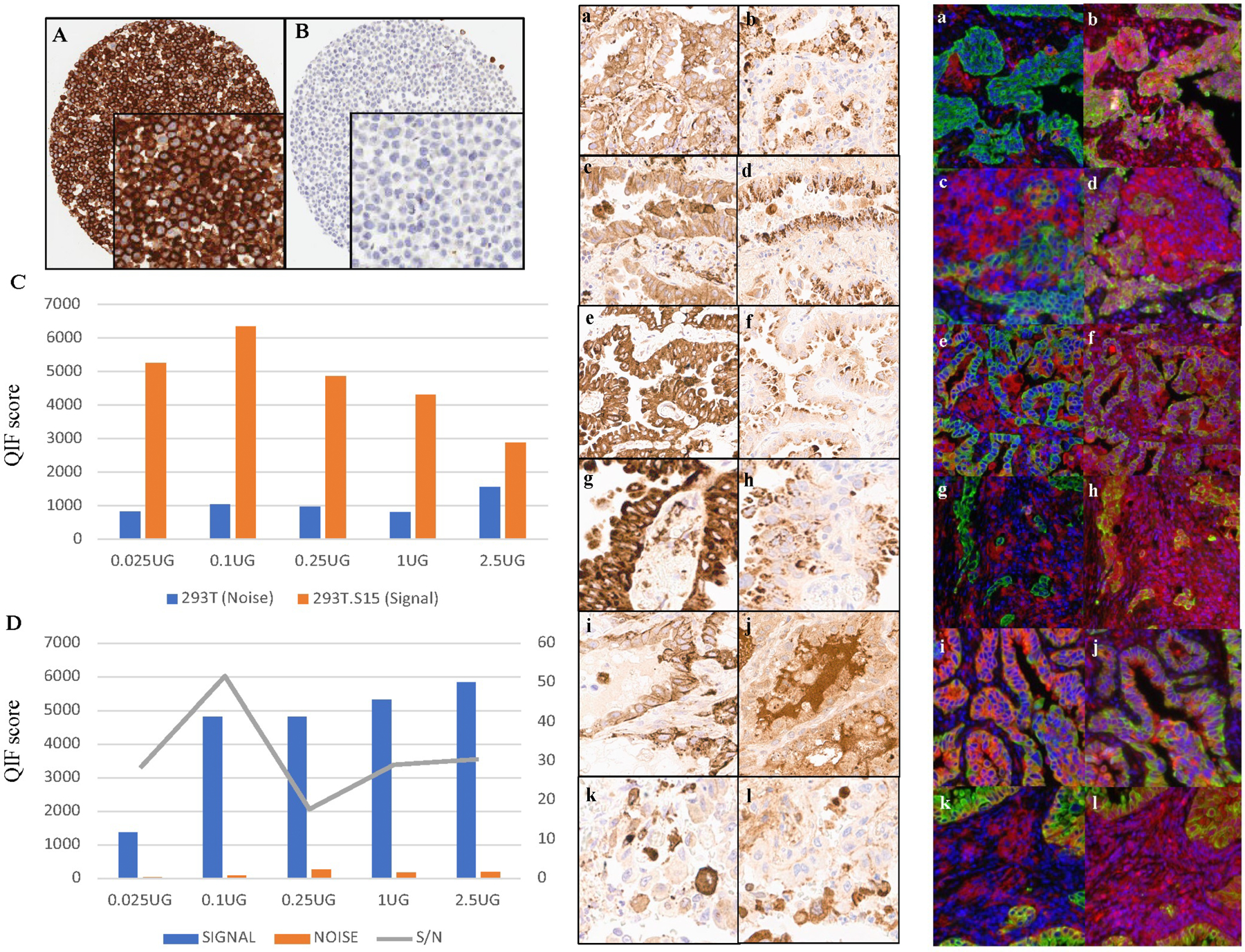
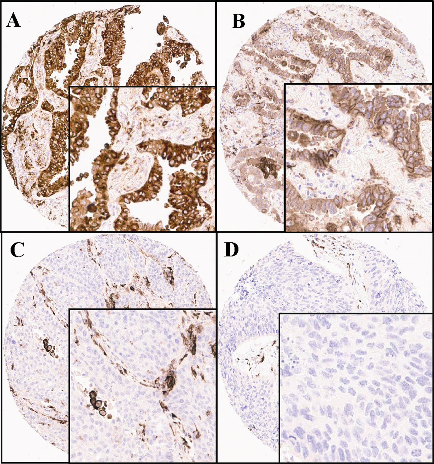
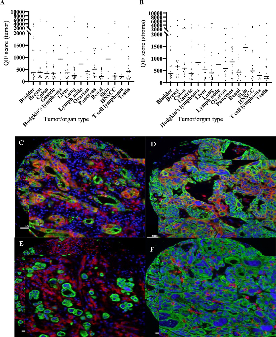
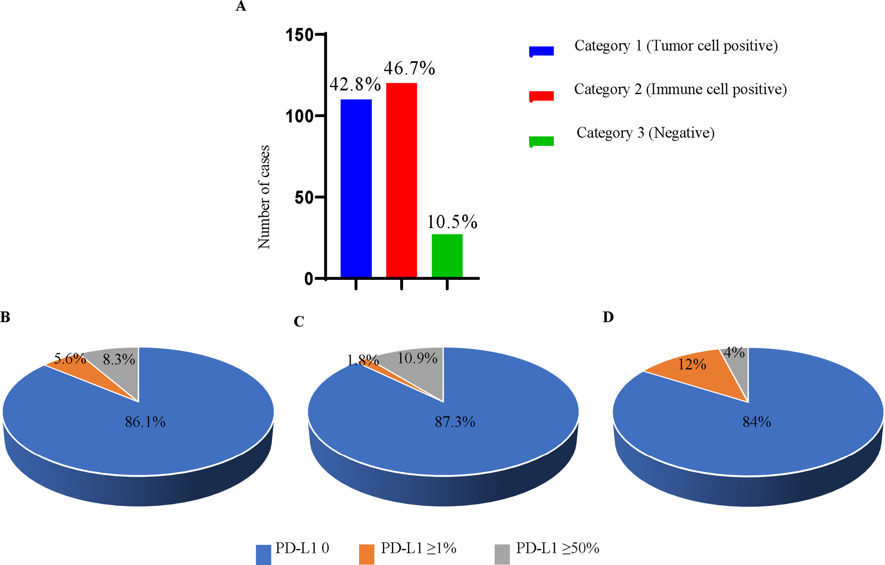
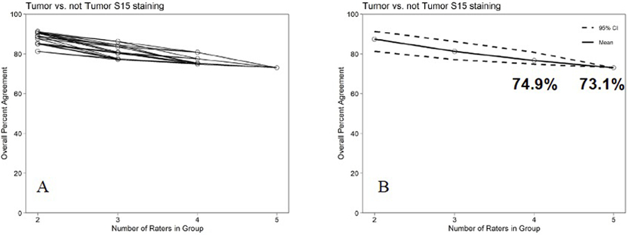
Siglec-15, a member of sialic-acid binding immunoglobulin type lectins, is normally expressed by myeloid cells and upregulated in some human cancers and represents a promising new target for immunotherapy. While PD-L1 blockade is an important strategy for immunotherapy, its effectiveness is limited. The expression of Siglec-15 has been demonstrated to be predominantly mutually exclusive to PD-L1 in certain cancer histologies. Thus, there is significant opportunity for Siglec-15 as an immunotherapeutic target for patients that do not respond to PD-1/PD-L1 inhibition. The aim of this study was to prospectively develop an immunohistochemical (IHC) assay for Siglec-15 to be used as a companion diagnostic for future clinical trials. Here, we create and validate an IHC assay with a novel recombinant antibody to the cytoplasmic domain of Siglec-15. To find an enriched target, this antibody was first used in a quantitative fluorescence (QIF) assay to screen a broad range of tumor histologies to determine tumor types where Siglec-15 demonstrated high expression. Based on this and previous data, we focused on development of a chromogenic IHC assay for lung cancer. Then we developed a scoring system for this assay that has high concordance amongst pathologist readers. We then use this chromogenic IHC assay to test the expression of Siglec-15 in two cohorts of NSCLC. We found that this assay shows a higher level of staining in both tumor and immune cells compared to previous QIF assays utilizing a polyclonal antibody. However, similar to that study, only a small percentage of positive Siglec-15 cases showed high expression for PD-L1. This validated assay for Siglec-15 expression may support development of a companion diagnostic assay to enrich for patients expressing the Siglec-15 target for therapy.


Conflict of interest statement

Competing Interests DLR has served as an advisor for AstraZeneca, Agendia, Amgen, BMS, Cell Signaling Technology, Cepheid, Daiichi Sankyo, Novartis, GSK, Konica Minolta, Merck, NanoString, PAIGE.AI, Perkin Elmer, Roche, Sanofi, Ventana and Ultivue. Amgen, Cepheid, Konica Minolta, NavigateBP, NextCure, and Lilly fund research in his lab. Sue Niu, Linda, Liu, Zac Cusumano and Sol Langermann are employees of NextCure. J. Zugazagoitia has served as a consultant for Astra Zeneca, BMS, Roche, Pfizer, Novartis, and Guardant Health. Reports speakers’ honoraria from BMS, Pfizer, Roche, Astra Zeneca, NanoString and Guardant Health. Reports travel honoraria from BMS, Pfizer, Roche, Astra Zeneca, and NanoString. Receives research support/funds from BMS, Astra Zeneca, and Roche.

Figures

Figure 1.
Figure 1.. Validation of Siglec-15 antibody.
A Membrane staining seen in the transfected cell line 293T.S15 (positive control), confirming the architectural localization of Siglec-15. B No Siglec-15 is seen in non-transfected cell line 293T (negative control). C Signal-to-noise plot for Siglec-15 in cell lines 293T (noise) and 293T.S15 (signal). D Signal-to-noise plot in tumor cores (YTMA 403) showing an optimal concentration at 0.1μg/ml. E, F Comparison of two clones of Siglec-15 in NSCLC by IHC (E) and QIF (F). The images on the left panel are stained with 1F7 (a, c, e, g, i, k). The images on the right are serial sections of the same spots stained with NP411 (b, d, f, h, j, l); red-Siglec-15, blue-DAPI (4′,6-diamidino-2-phenylindole), green-Cytokeratin.
Figure 2.
Figure 2.
Ordinal (binary) Scoring System for Siglec-15 with Category 1, which shows staining in at least 10% of tumor cells regardless of intensity (A, B), Category 2 with staining in at least 10% of immune cells (C) and Category 3 with no evidence of any staining (D).
Figure 3.
Figure 3.. Expression of Siglec-15 in different cancers by immunofluorescence.
Expression of Siglec-15 in different cancers by immunofluorescence. A, B Lung, bladder, and breast cancers are seen to express high Siglec-15 seen as high QIF scores by AQUA in tumor (A) and stroma (B). Representative images of Siglec-15 expression (red) in tumor and stroma in lung (C), predominantly in tumor cells in bladder cancer (D), mainly in stromal immune cells in bladder cancer (E) and predominantly in stromal immune cells in breast cancer (F); red-Siglec-15, blue-DAPI (4′,6-diamidino-2-phenylindole), green-Cytokeratin.
Figure 4.
Figure 4.
Comparison of Siglec-15 and PD-L1 in tumor showed some-what mutually exclusive expression. A Overall expression of Siglec-15 in NSCLC (YTMA 423). B PD-L1 expression in Category 1 (tumor cell positive for Siglec-15). C PD-L1 in Category 2 (immune cell positive for Siglec-15). D PD-L1 in Category 3 (no staining for Siglec-15).
Figure 5.
Figure 5.. ONEST plots for assessment of tumor staining of Siglec 15.
A. The OPA for set of 2 to 5 raters for tumor staining by Siglec 15. B. The mean and 95% lower bound for the OPAs for 2 to 5 observers.
Figure 6.
Figure 6.. Siglec-15 in different mutation types of NSCLC (YTMA 310).
A Siglec-15 expression in Category 1 (tumor cell positive for Siglec-15). B Siglec-15 expression in Category 2 (immune cell positive for Siglec-15). C Siglec-15 expression in Category 3 (no positive

References

    1. Crocker PR &Varki, A. Siglecs, sialic acids and innate immunity. Trends Immunol 22, 337–342 (2001). - PubMed
    1. Adams OJ, Stanczak MA, von Gunten S & Laubli H Targeting sialic acid-Siglec interactions to reverse immune suppression in cancer. Glycobiology 28, 640–647 (2018). - PubMed
    1. Varki A & Angata T Siglecs--the major subfamily of I-type lectins. Glycobiology 16, 1R–27R (2006). - PubMed
    1. Wang J, Sun J, Liu LN, Flies DB, Nie X, Toki M et al. Siglec-15 as an immune suppressor and potential target for normalization cancer immunotherapy. Nat Med 25, 656–666 (2019). - PMC - PubMed
    1. Angata T, Tabuchi Y, Nakamura K &Nakamura, M. Siglec-15: an immune system Siglec conserved throughout vertebrate evolution. Glycobiology 17, 838–846 (2007). - PubMed

Publication types

MeSH terms

Substances