

SARS-CoV-2 vaccination can elicit a CD8 T-cell dominant hepatitis
- PMID: 35461912
- PMCID: PMC9021033
- DOI: 10.1016/j.jhep.2022.03.040
SARS-CoV-2 vaccination can elicit a CD8 T-cell dominant hepatitis
Abstract
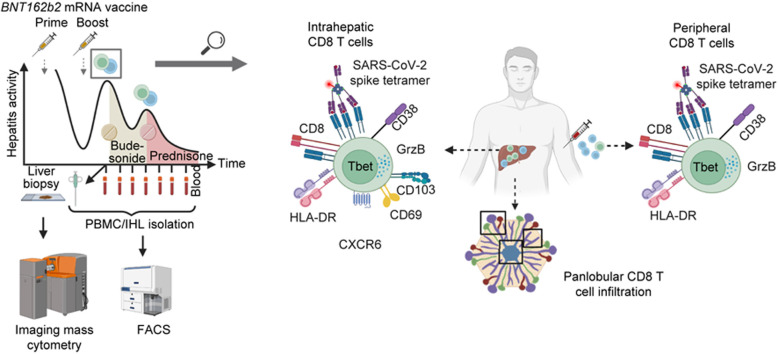
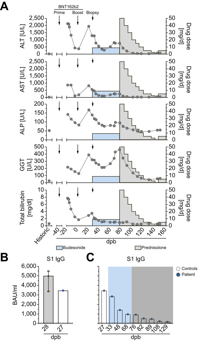
Background & aims: Autoimmune hepatitis episodes have been described following SARS-CoV-2 infection and vaccination but their pathophysiology remains unclear. Herein, we report the case of a 52-year-old male, presenting with bimodal episodes of acute hepatitis, each occurring 2-3 weeks after BNT162b2 mRNA vaccination. We sought to identify the underlying immune correlates. The patient received oral budesonide, relapsed, but achieved remission under systemic steroids.
Methods: Imaging mass cytometry for spatial immune profiling was performed on liver biopsy tissue. Flow cytometry was performed to dissect CD8 T-cell phenotypes and identify SARS-CoV-2-specific and EBV-specific T cells longitudinally. Vaccine-induced antibodies were determined by ELISA. Data were correlated with clinical laboratory results.
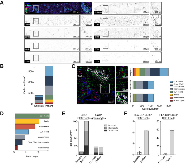
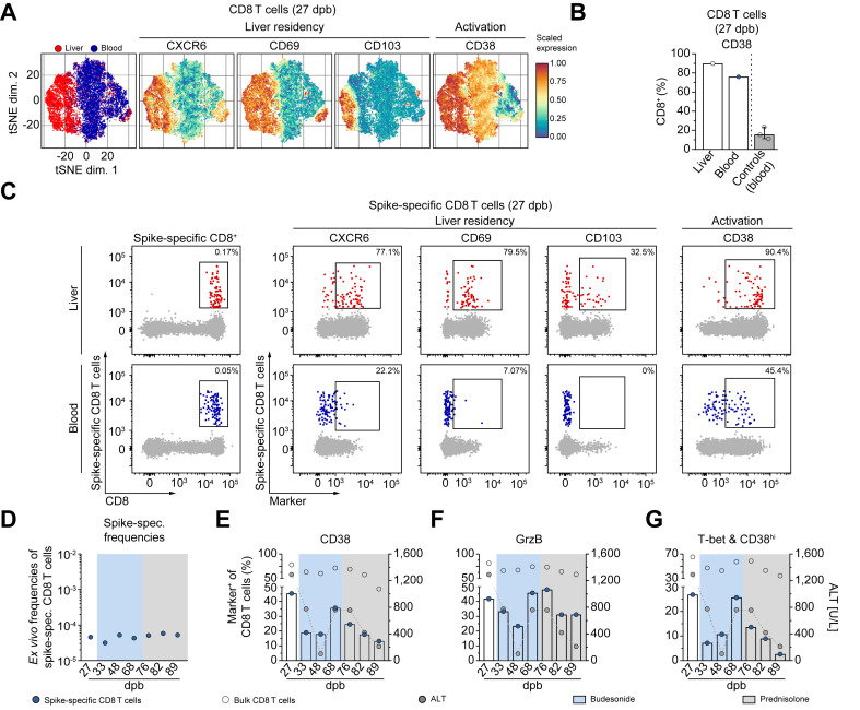
Results: Analysis of the hepatic tissue revealed an immune infiltrate quantitatively dominated by activated cytotoxic CD8 T cells with panlobular distribution. An enrichment of CD4 T cells, B cells, plasma cells and myeloid cells was also observed compared to controls. The intrahepatic infiltrate showed enrichment for CD8 T cells with SARS-CoV-2-specificity compared to the peripheral blood. Notably, hepatitis severity correlated longitudinally with an activated cytotoxic phenotype of peripheral SARS-CoV-2-specific, but not EBV-specific, CD8+ T cells or vaccine-induced immunoglobulins.
Conclusions: COVID-19 vaccination can elicit a distinct T cell-dominant immune-mediated hepatitis with a unique pathomechanism associated with vaccination-induced antigen-specific tissue-resident immunity requiring systemic immunosuppression.
Lay summary: Liver inflammation is observed during SARS-CoV-2 infection but can also occur in some individuals after vaccination and shares some typical features with autoimmune liver disease. In this report, we show that highly activated T cells accumulate and are evenly distributed in the different areas of the liver in a patient with liver inflammation following SARS-CoV-2 vaccination. Moreover, within the population of these liver-infiltrating T cells, we observed an enrichment of T cells that are reactive to SARS-CoV-2, suggesting that these vaccine-induced cells can contribute to liver inflammation in this context.
Keywords: Autoimmune hepatitis; CD8+ T cell; COVID-19; Immunosuppression; Vaccination; Virus-specific T cell.
Copyright © 2022 European Association for the Study of the Liver. Published by Elsevier B.V. All rights reserved.
Conflict of interest statement
Conflict of interest The authors declare no conflicts of interest that pertain to this work. Please refer to the accompanying ICMJE disclosure forms for further details.
Figures
Comment in
-
Immune-mediated liver injury represented as overlap syndrome after SARS-CoV-2 vaccination.J Hepatol. 2022 Oct;77(4):1209-1211. doi: 10.1016/j.jhep.2022.06.029. Epub 2022 Jul 8. J Hepatol. 2022. PMID: 35817223 Free PMC article. No abstract available.
-
In situ detection of vaccine mRNA in the cytoplasm of hepatocytes during COVID-19 vaccine-related hepatitis.J Hepatol. 2023 Jan;78(1):e20-e22. doi: 10.1016/j.jhep.2022.08.039. Epub 2022 Sep 15. J Hepatol. 2023. PMID: 36116717 Free PMC article. No abstract available.
References
-
- Clayton-Chubb D., Schneider D., Freeman E., Kemp W., Roberts S.K. Comment to the letter of Bril F et al. “Autoimmune hepatitis developing after coronavirus disease 2019 (COVID-19) vaccine: causality or casualty?”. J Hepatol. 2021
Publication types
MeSH terms
Substances
LinkOut - more resources
Full Text Sources
Other Literature Sources
Medical
Research Materials
Miscellaneous
