Successful Conservative Treatment of an Acute Arterial Occlusion After Total Knee Arthroplasty: Report of 2 Cases and Review of the Literature
- PMID: 35462393
- PMCID: PMC9047693
- DOI: 10.12659/AJCR.936295
Successful Conservative Treatment of an Acute Arterial Occlusion After Total Knee Arthroplasty: Report of 2 Cases and Review of the Literature
Abstract
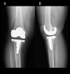
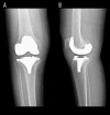
BACKGROUND Acute arterial occlusion after total knee arthroplasty (TKA) is a rare but occasionally limb-threatening complication. Successful outcomes of surgical treatment for acute arterial occlusion after TKA have been frequently reported in the literature; however, few reports have described conservative treatment. This case report describes the successful conservative treatment of popliteal artery occlusion after TKA. CASE REPORT We report 2 cases of popliteal artery occlusion after TKA that were managed with conservative treatment. In Case 1, a 68-year-old woman presented with a weak dorsalis pedis pulse in the foot and weakness to dorsiflexion of the toe on the operative side immediately after TKA. The operative lower extremity arterial ultrasonography and computed tomography angiography demonstrated the popliteal artery occlusion. In Case 2, a 79-year-old woman presented a cold right foot and lack of popliteal and dorsalis pedis pulse in the operated extremity immediately after TKA, and Doppler ultrasound did not reveal a flow for the dorsalis pedis artery. In both patients, urgent angiographies showed popliteal artery occlusion, and blood flow was observable in the anterior tibial, peroneal, and foot arteries collateral perfusion. Thus, conservative treatments were chosen, and anticoagulant and vasodilator therapies were undergone in both patients. At 6 months after surgery, they were able to walk without intermittent claudication. CONCLUSIONS Conservative treatment can be a good option for popliteal artery occlusion after TKA in cases of rich collateral circulation.
Conflict of interest statement
Figures


References
-
- Jones CA, Beaupre LA, Johnston DW, Suarez-Almazor ME. Total joint arthroplasties: Current concepts of patient outcomes after surgery. Clin Geriatr Med. 2005;21(3):527–41. , vi. - PubMed
-
- Menendez ME, Memtsoudis SG, Opperer M, et al. A nationwide analysis of risk factors for in-hospital myocardial infarction after total joint arthroplasty. Int Orthop. 2015;39(4):777–86. - PubMed
-
- Matziolis G, Perka C, Labs K. Acute arterial occlusion after total knee arthroplasty. Arch Orthop Trauma Surg. 2004;124(2):134–36. - PubMed
-
- Rand JA. Vascular complications of total knee arthroplasty. Report of three cases. J Arthroplasty. 1987;2(2):89–93. - PubMed
Publication types
MeSH terms
LinkOut - more resources
Full Text Sources
Medical

