

Tuberculosis detection in chest radiograph using convolutional neural network architecture and explainable artificial intelligence
- PMID: 35462630
- PMCID: PMC9016694
- DOI: 10.1007/s00521-022-07258-6
Tuberculosis detection in chest radiograph using convolutional neural network architecture and explainable artificial intelligence
Abstract
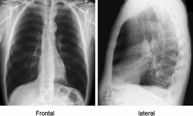
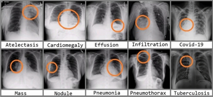
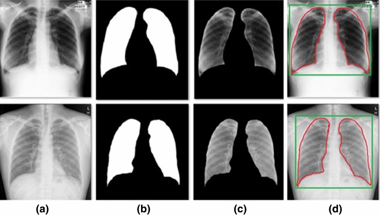
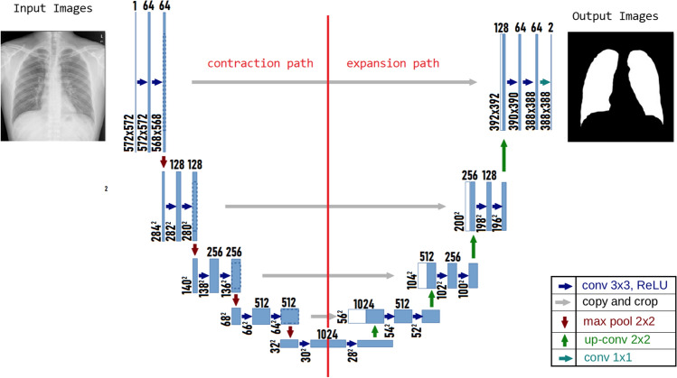
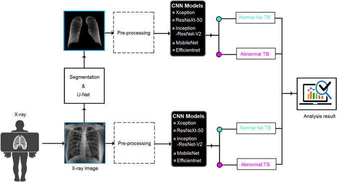
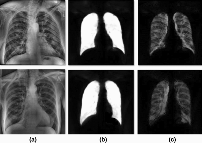
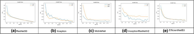
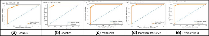
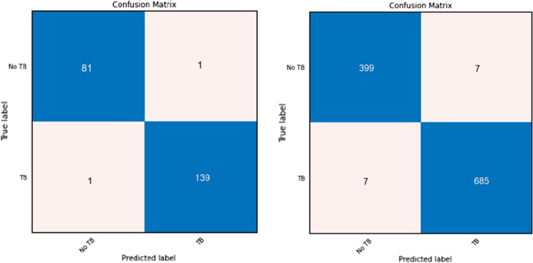
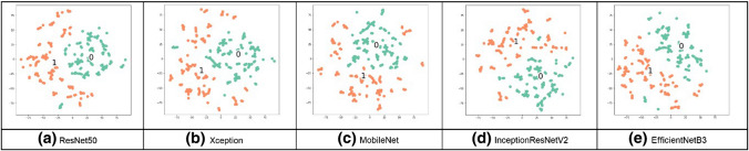
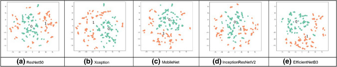
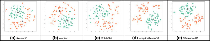
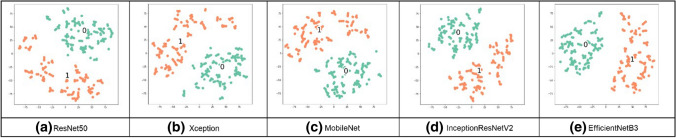
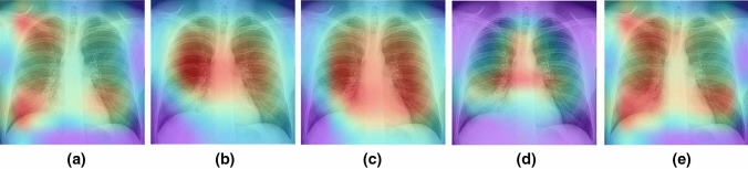
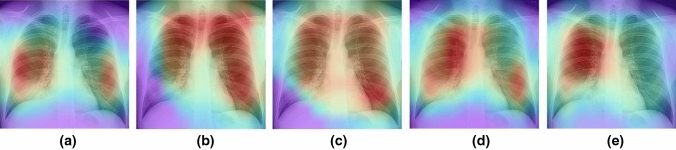
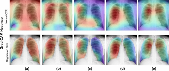
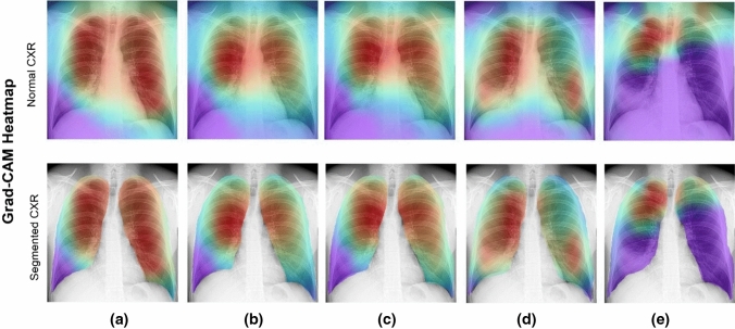
In most regions of the world, tuberculosis (TB) is classified as a malignant infectious disease that can be fatal. Using advanced tools and technology, automatic analysis and classification of chest X-rays (CXRs) into TB and non-TB can be a reliable alternative to the subjective assessment performed by healthcare professionals. Thus, in the study, we propose an automatic TB detection system using advanced deep learning (DL) models. A significant portion of a CXR image is dark, providing no information for diagnosis and potentially confusing DL models. Therefore, in the proposed system, we use sophisticated segmentation networks to extract the region of interest from multimedia CXRs. Then, segmented images are fed into the DL models. For the subjective assessment, we use explainable artificial intelligence to visualize TB-infected parts of the lung. We use different convolutional neural network (CNN) models in our experiments and compare their classification performance using three publicly available CXR datasets. EfficientNetB3, one of the CNN models, achieves the highest accuracy of 99.1%, with a receiver operating characteristic of 99.9%, and an average accuracy of 98.7%. Experiment results confirm that using segmented lung CXR images produces better performance than does using raw lung CXR images.
Keywords: Chest X-Ray; Convolution neural networks; Deep learning; Image segmentation; Tuberculosis detection.
© The Author(s), under exclusive licence to Springer-Verlag London Ltd., part of Springer Nature 2022.
Conflict of interest statement
Conflict of interestThe authors declare that they have no known competing financial interests or personal relationships that could have appeared to influence the work reported in this paper.
Figures
References
-
- Haloi M, Rajalakshmi KR, Walia P (2018) Towards radiologist-level accurate deep learning system for pulmonary screening, arXiv:1807.03120 [cs.CV]
-
- Chandra TB, et al. Automatic detection of Tuberculosis related abnormalities in chest Xray images using hierarchical feature extraction scheme. Expert Syst Appl. 2020;158(15):113514. doi: 10.1016/j.eswa.2020.113514. - DOI
-
- Hooda R, Sofat S, Kaur S, Mittal A, (2017) Deep-learning: a potential method for tuberculosis detection using chest radiography. In: 2017 IEEE international conference on signal and image processing applications (ICSIPA), Kuching, pp. 497–502
-
- Liu et al., (2017), “TX-CNN: Detecting tuberculosis in chest X-ray images using convolutional neural network. 2017 IEEE international conference on image processing (ICIP), Beijing, pp. 2314–2318
LinkOut - more resources
Full Text Sources