. 2022 Apr 6:13:861938.
doi: 10.3389/fphar.2022.861938. eCollection 2022.

mPEG-PDLLA Micelles Potentiate Docetaxel for Intraperitoneal Chemotherapy in Ovarian Cancer Peritoneal Metastasis

Affiliations

mPEG-PDLLA Micelles Potentiate Docetaxel for Intraperitoneal Chemotherapy in Ovarian Cancer Peritoneal Metastasis

Yumei Zhang et al. Front Pharmacol. .

Abstract

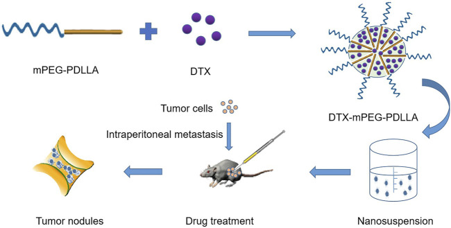
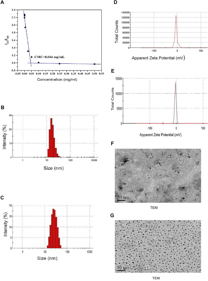
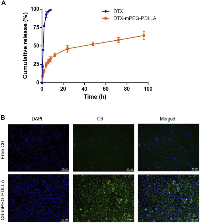
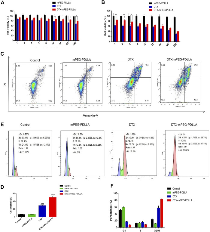
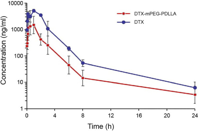
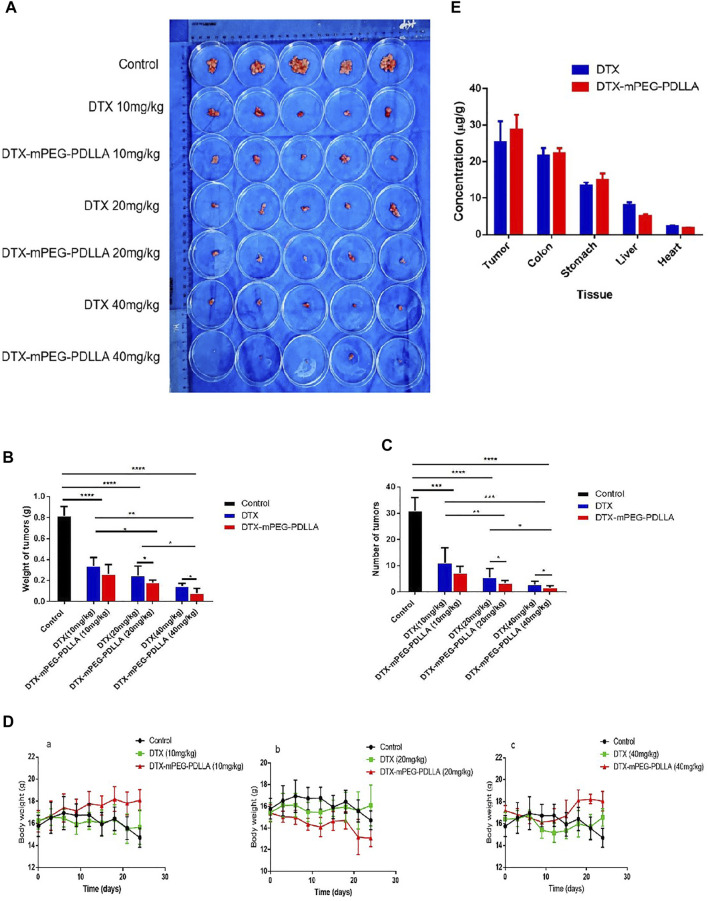
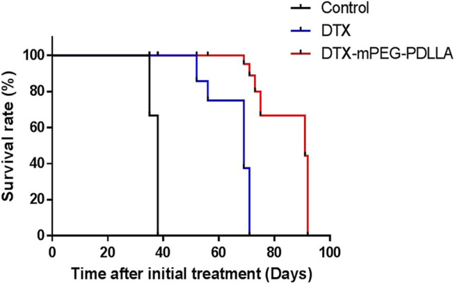
Ovarian cancer is the second most common cause of gynecological cancer death in women. It is usually diagnosed late and accompanied by peritoneal metastasis. For ovarian cancer with peritoneal metastasis, intraperitoneal (IP) chemotherapy can maintain a high drug concentration in the abdominal cavity and reduce local and systemic toxicity. Recently, docetaxel (DTX) has shown broad-spectrum antitumor activity against various malignant tumors, including ovarian cancer with peritoneal metastasis. However, DTX has limited clinical applications due to its poor water solubility, predisposition to hypersensitivity, fluid retention, and varying degrees of neurotoxicity. In this study, we prepared methoxy-poly(ethylene glycol)-block-poly(D,L-lactide) (mPEG-PDLLA) micelles loaded with DTX and developed an alternative, less toxic, more effective DTX formulation, without Tween 80, and evaluated its pharmacokinetics in the abdominal cavity and its efficacy in ovarian cancer with peritoneal metastasis. The mean diameter of DTX-mPEG-PDLLA was about 25 nm, and the pharmacokinetics of BALB/c mice via IP showed that the plasma exposure of DTX-mPEG-PDLLA was about four times lower than that of DTX. Importantly, DTX-mPEG-PDLLA was significantly more effective than DTX and prolonged the survival period in a SKOV-3 ovarian cancer peritoneal metastasis model. Moreover, the apoptosis rate was significantly increased in vitro. Based on these findings, it is expected that DTX-mPEG-PDLLA can enhance efficacy against ovarian cancer peritoneal metastasis, while reducing toxic side effects, and has the potential to be used in the clinical treatment of peritoneal metastatic cancer.

Keywords: DTX-mPEG-PDLLA micelles; intraperitoneal administration; ovarian cancer; peritoneal metastasis; pharmacokinetics.


Conflict of interest statement

The authors declare that the research was conducted in the absence of any commercial or financial relationships that could be construed as a potential conflict of interest.

Figures

FIGURE 1
FIGURE 1
Schematic of the synthesis and intraperitoneal administration of DTX-mPEG-PDLLA micelles. The DTX-mPEG-PDLLA micelles were prepared with DTX and mPEG-PDLLA polymers by a self-assembly method. The drugs were delivered by the micelles after IP administration for an antitumor effect in vivo.
FIGURE 2
FIGURE 2
Characterization of DTX-mPEG-PDLLA micelles. (A) CMC measurement of mPEG-PDLLA by a pyrene fluorescence probe. (B,C) Particle size distribution of mPEG-PDLLA and DTX-mPEG-PDLLA micelles. (D,E) Zeta potential of mPEG-PDLLA and DTX-mPEG-PDLLA micelles. (F,G) TEM images of mPEG-PDLLA and DTX-mPEG-PDLLA micelles.
FIGURE 3
FIGURE 3
DTX release from DTX-mPEG-PDLLA micelles in vitro and cellular uptake of C6-mPEG-PDLLA micelles. (A) Release characteristics of DTX and DTX-mPEG-PDLLA micelles in vitro. The data were analyzed as mean ± SD (n = 3). (B) Cellular uptake in SKOV-3 cells.
FIGURE 4
FIGURE 4
Antitumor activity of DTX-mPEG-PDLLA micelles in ovarian cancer cells. (A,B) Cytotoxicity of mPEG-PDLLA, DTX, and DTX-mPEG-PDLLA micelles in SKOV-3 human ovarian cancer cells treated for 24 and 48 h, respectively (n = 3, mean ± SD); *p < 0.05 and **p < 0.01. (C) Apoptosis induction of SKOV-3 cells treated with mPEG-PDLLA, DTX, and DTX-mPEG-PDLLA for 48 h analyzed by the Annexin V-FITC Apoptosis Detection Kit. (D) Results representing three-independent experiments; ***p < 0.001, ****p < 0.0001 vs. the control group. (E,F) Cell cycle assays and analysis of mPEG-PDLLA, DTX, and DTX-mPEG-PDLLA micelles in SKOV-3 cells treated for 48 h, respectively (n = 3, mean ± SD).
FIGURE 5
FIGURE 5
Pharmacokinetics of DTX-mPEG-PDLLA micelles by IP administration in vivo. Plasma concentration–time curves of free DTX and DTX-mPEG-PDLLA micelles in mice via IP administration (n = 6).
FIGURE 6
FIGURE 6
Antitumor effect and tissue distribution of DTX-mPEG-PDLLA micelles by IP administration in ovarian cancer peritoneal metastasis. (A) Photographs of peritoneal cavity tumors in each group on day 24 after IP administration. (B) Analysis of tumor nodules weight. (C) Numbers of tumor nodules. (D) Changes in body weight of mice in the 10 mg/kg (a), 20 mg/kg groups (b), 40 mg/kg groups (c), and the control group, which was injected with NS by IP administration. (E) Comparison of DTX and DTX-mPEG-PDLLA in tissues and tumors 1 h by IP administration at 10 mg/kg dosage. *p < 0.05, **p < 0.01, ***p < 0.001, and ****p < 0.0001.
FIGURE 7
FIGURE 7
Survival analysis of DTX-mPEG-PDLLA micelles via IP administration in ovarian cancer peritoneal metastasis. Survival curves for SKOV-3 tumor-bearing nude mice receiving different treatments. Mice were treated with NS, DTX, or DTX-mPEG-PDLLA micelles on days 1 and 7.

Similar articles

Cited by

References

    1. Armstrong D. K., Bundy B., Winzel L., Huang H. Q., Baergen R., Lele S., et al. (2006). Intraperitoneal Cisplatin and Paclitaxel in Ovarian Cancer. Obstetrical Gynecol. Surv. 61, 240–242. 10.1097/01.ogx.0000206353.22975.c5 - DOI - PubMed
    1. Byagari K., Shanavas A., Rengan A. K., Kundu G. C., Srivastava R. (2014). Biocompatible Amphiphilic Pentablock Copolymeric Nanoparticles for Anti-Cancer Drug Delivery. J. Biomed. Nanotechnol. 10, 109–119. 10.1166/jbn.2014.1791 - DOI - PubMed
    1. Bray F., Ferlay J., Soerjomataram I., Siegel R. L., Torre L. A., Jemal A. (2018). Global Cancer Statistics 2018: GLOBOCAN Estimates of Incidence and Mortality Worldwide for 36 Cancers in 185 Countries. CA. Cancer J. Clin. 68, 394–424. 10.3322/caac.21492 - DOI - PubMed
    1. Chen J., Jiang Z., Zhang Y. S., Ding J., Chen X. (2021). Smart Transformable Nanoparticles for Enhanced Tumor Theranostics. Appl. Phys. Rev. 8, 041321. 10.1063/5.0061530 - DOI
    1. de Bree E., Rosing H., Michalakis J., Romanos J., Relakis K., Theodoropoulos P. A., et al. (2006). Intraperitoneal Chemotherapy with Taxanes for Ovarian Cancer with Peritoneal Dissemination. Eur. J. Surg. Oncol. 32, 666–670. 10.1016/j.ejso.2006.03.008 - DOI - PubMed

LinkOut - more resources