. 2022 Apr 20:13:20417314221091033.
doi: 10.1177/20417314221091033. eCollection 2022 Jan-Dec.

Toward 3D-bioprinting of an endocrine pancreas: A building-block concept for bioartificial insulin-secreting tissue

Affiliations

Toward 3D-bioprinting of an endocrine pancreas: A building-block concept for bioartificial insulin-secreting tissue

Gabriel A Salg et al. J Tissue Eng. .

Abstract

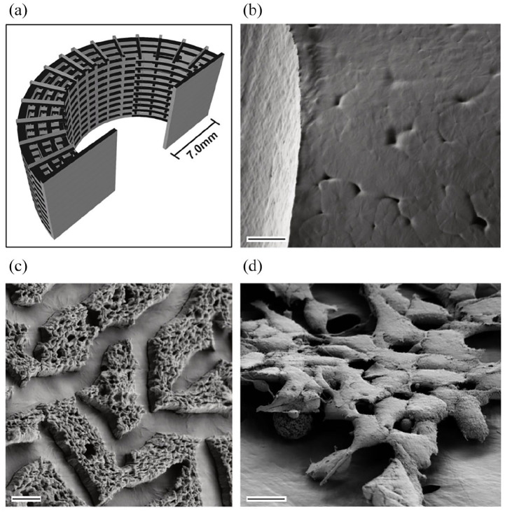
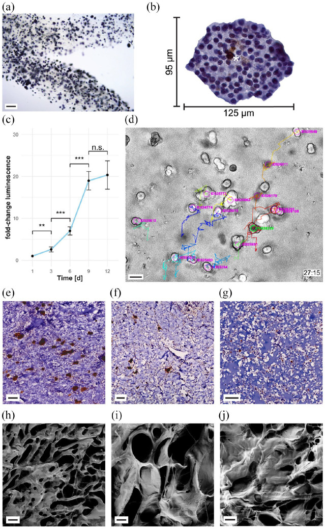
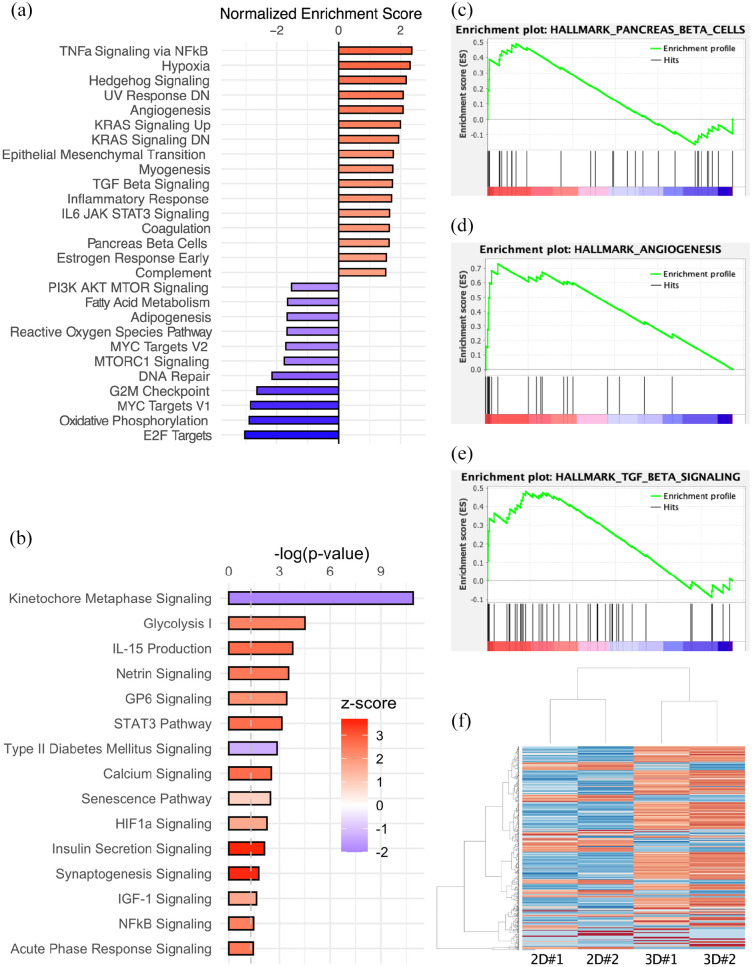
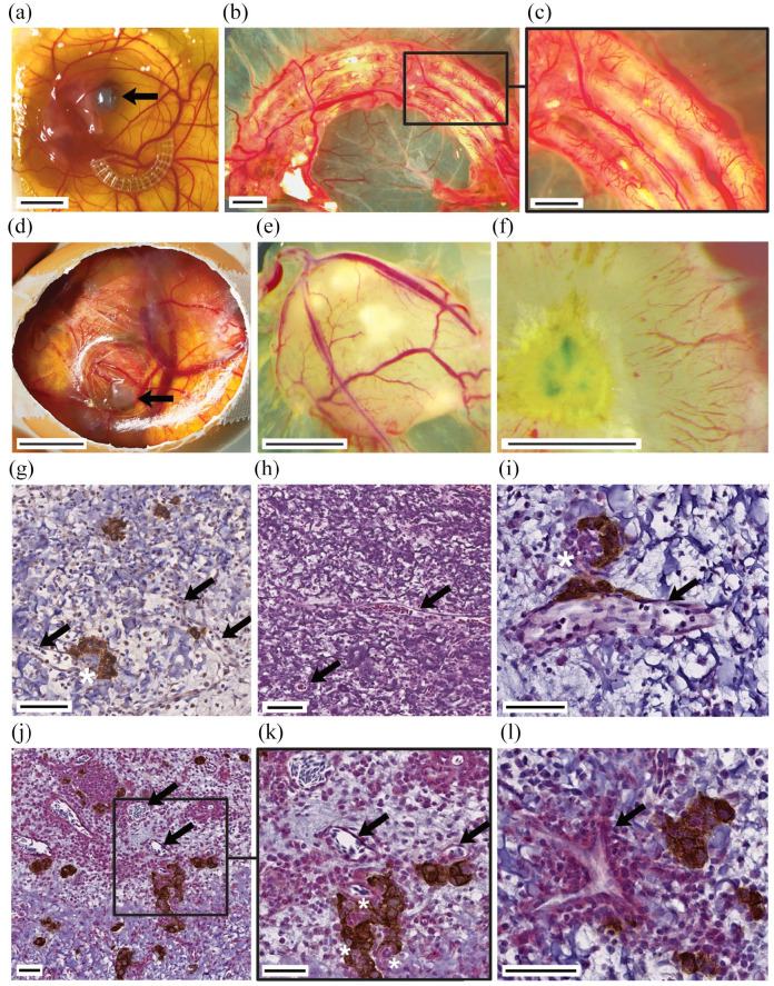
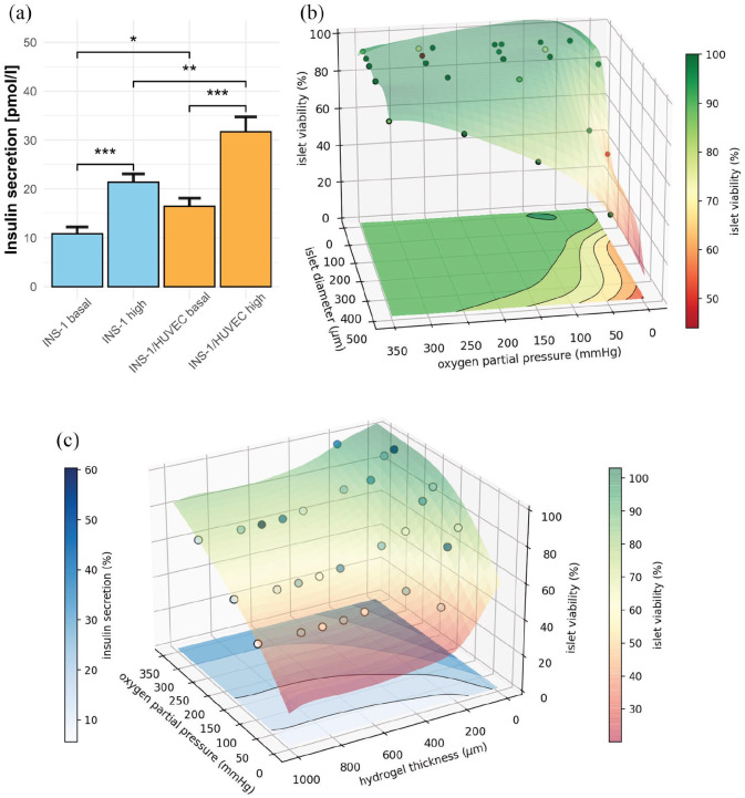
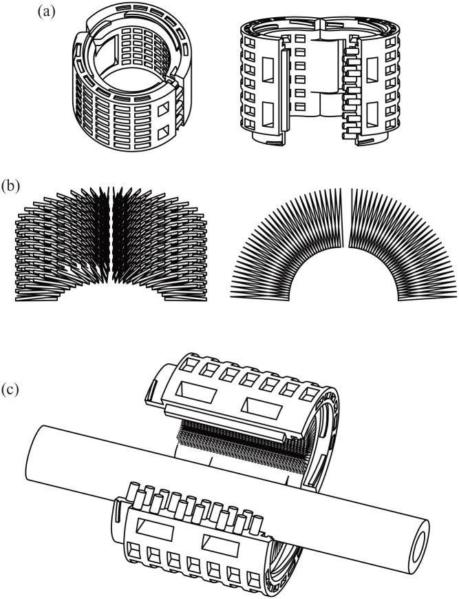
Three-dimensional bioprinting of an endocrine pancreas is a promising future curative treatment for patients with insulin secretion deficiency. In this study, we present an end-to-end concept from the molecular to the macroscopic level. Building-blocks for a hybrid scaffold device of hydrogel and functionalized polycaprolactone were manufactured by 3D-(bio)printing. Pseudoislet formation from INS-1 cells after bioprinting resulted in a viable and proliferative experimental model. Transcriptomics showed an upregulation of proliferative and ß-cell-specific signaling cascades, downregulation of apoptotic pathways, overexpression of extracellular matrix proteins, and VEGF induced by pseudoislet formation and 3D-culture. Co-culture with endothelial cells created a natural cellular niche with enhanced insulin secretion after glucose stimulation. Survival and function of pseudoislets after explantation and extensive scaffold vascularization of both hydrogel and heparinized polycaprolactone were demonstrated in vivo. Computer simulations of oxygen, glucose and insulin flows were used to evaluate scaffold architectures and Langerhans islets at a future perivascular transplantation site.

Keywords: Bioprinting; diabetes; endocrine pancreas; next-generation sequencing; tissue engineering.


Conflict of interest statement

Declaration of conflicting interests: The author(s) declared the following potential conflicts of interest with respect to the research, authorship, and/or publication of this article: G.A.S., T.H. and H.G.K. are named inventors of a European patent application of University Hospital Heidelberg. Other authors declare that no competing financial interests exist.

Figures

Figure 1.
Figure 1.
Solid polymer scaffold component was functionalized to enhance applicability, cell adhesion, and vascular ingrowth. (a) CAD model of PCL scaffold for 3D-printing as used in the experiments. (b) Scanning electron microscopy of untreated PCL scaffold structure after 3D-printing shows smooth surface with minimal porosity. (c) Surface modification of 3D-printed PCL scaffold with heparin. (d) Adhesion of INS-1 on 3D-printed PCL scaffold. Scale bar (b) 20 µm, (c) 2 µm, and (d) 10 µm.
Figure 2.
Figure 2.
Hydrogel structure initiates pseudoislet formation. 3D-bioprinted INS-1 cells remain viable, migrate and proliferate. (a) MTT assay of pseudoislets formed after bioprinting (day 5 post-printing) of INS-1 containing gelatin methacrylate hydrogel shows metabolically active pseudoislets spatially distributed in 3D matrix. For further data s. Supplementary Figure 3 (b) Immunohistochemical staining of cleaved caspase-3 (brown, asterisk). Depicted: Pseudoislet formed from bioprinted INS-1 cells cultivated for 12 days. Immunohistochemistry revealed few apoptotic cells in the core of the pseudoislet. Apoptotic single cells disseminated in the hydrogel structure could not be detected. For further data s. Supplementary Figure 4 (c) Proliferation assay of bioprinted cells. A plateau is reached after 9 days in culture, indicating maximum loading capacity. Error bars depict SEM, n ⩾ 13/timepoint. (d) Cell movement tracking of time lapse microscopical investigation with TrackMate. Colored movement tracks (27:15h after start of experiment) depict, that cells encapsulated in hydrogel, bioprinted, and 2 s UV-cured (here: day 3 post-printing) are able to migrate within the hydrogel. Live microscopy was performed at 328 time points with an interval of 5 min. For further data s. Supplementary Movie 2. (e–g) Immunohistochemical anti-insulin staining (brown areas) of CAM assay explants of bioprinted INS-containing hydrogel (e) after 2 s of UV curing, (f) 5 s of UV curing, and (g) 15 s of UV curing. In INS-containing hydrogel with 2 s crosslinking time (e) most insulin-stained areas were detected and pseudoislet formation is found. Spatially distributed pseudoislets remained viable and functional. Longer crosslinking periods (5 s (f), 15 s (g) showed few pseudoislets (f) or scattered INS-1 single cells (g). (h–j) Scanning electron microscopy of freeze-dried hydrogel structures (GelXA LAMININK 411) with different UV-curing times. Electron microscopy showed different morphological features of the hydrogel depending on the crosslinking time of (h) 2 s, (i) 5 s, (j) 10 s. The hydrogel micro-structure showed a homogenous porous structure (h). For further data s. Supplementary Figure 5A–C. Scale bar (a) 200 µm, (d) 20 µm, (e–g) 50 µm, and (h–j) 10 µm.
Figure 3.
Figure 3.
mRNA sequencing of 3D-bioprinted domes compared with monolayer culture control shows robust differential gene expression clusters and alteration of hallmark pathways. (a) Gene set enrichment analysis revealed significantly altered hallmark pathways (p < 0.05). (b) IPA revealed significantly altered canonical pathways (p < 0.05) and color-coded z-score indicates activation of pathways including insulin secretion signaling. Upregulated hallmark pathways include pancreas ß-cells (c), TGFß signaling (d), and angiogenesis (e). (f) Heatmap of alteration in gene enrichment (500 most significant alterations) comparing 3D-bioprinted culture with monolayer culture.
Figure 4.
Figure 4.
Chorioallantoic membrane assay is a suitable model for investigating angiogenesis in tissue-engineered grafts. Extensive, rapid vascular ingrowth is seen in both PCL and cell-laden hydrogel structure after the 9-day assay period. Ex ovo CAM assay experiments enabled direct comparison of heparinized PCL scaffolds with untreated controls and validated the beneficial properties of heparinization for enhanced vascular ingrowth ((a–c) arrow indicates eye of chicken embryo). In ovo CAM assay experiments were used for investigation of 3D-bioprinted INS-1-laden droplets ((d–l) arrow indicates xenotransplant). Vascular structures (arrows) penetrated into the scaffold (g–l). (g) Anti-insulin immunohistochemical staining of CAM assay explant. (h) H&E staining of CAM assay explant. (i–l) Anti-insulin (brown) and anti-CD31 (red) immunohistochemical double-staining of CAM assay explant. Rapid vascularization maintained viability and function of pseudoislets. Peri- and intra-insular (asterisks) vessels were detected (g–l). Scale bar (a and d) 10 mm, (b, c, e, and f) 2 mm, (j) 100 µm, (g, h, k, and l) 50 µm, (i) 20 µm.
Figure 5.
Figure 5.
Insulin-secretion of bioprinted INS-1 cells is enhanced in co-culture with EC. Computer simulation of human Langerhans islets predicts concept feasibility and defines boundary conditions for viability and function of bioprinted 3D hydrogel geometries. (a) Insulin secretion of hydrogel-embedded INS-1 culture and INS-1/HUVEC co-culture at basal and high glucose levels 3 days post printing. 3D-Bioprinted, encapsulated INS-1 cells are responsive to glucose stimulation. The INS-1/HUVEC co-culture ameliorates the amount of insulin secreted. Insulin levels for both experimental settings were normalized to 10,000 cells. Error bars depict SEM, n ⩾ 20/condition. (b) Simulation of viability of human islets of Langerhans encapsuled in hydrogel by finite element analysis. Calculation for 400 µm constant diffusion distance through the hydrogel. Glucose inflow concentration is kept at 10 mM. Inflow oxygen partial pressure ranging from 5 mmHg to 350 mmHg, islet diameter from 100 µm to 500 µm. Circles represent data points, gathered by diffusion simulation, and colored semitransparent surface and projection on the base represent fit through cell viability data. (c) Simulation of insulin secretion and viability. Constant glucose concentration of 10 mM, constant islet diameter of 500 µm, inflow oxygen partial pressure varying between 50 mmHg and 350 mmHg, hydrogel shell thickness ranging from 0 µm to 1000 µm. Circles represent calculated datapoints, semitransparent surface represents fit through 3D cell viability (right color bar). Insulin secretion is displayed as contour lines at the bottom of the diagram (left color bar). Simulations were performed for 464 different scenarios.
Figure 6.
Figure 6.
Proposed application concept of a perivascular implant based on a hybrid scaffold building-blocks. (a) CAD-drawing of polycaprolactone shell component: Proposed 3D-printed outer layer. (b) CAD-drawing of cell-encapsulating hydrogel structure: Proposed bioprinted inner layer. (c) CAD-drawing of proposed merged hybrid perivascular device for future application in human. In this study, building-blocks for a future insulin-producing perivascular implant were investigated in vitro and in ovo. In further studies in vivo, we propose merging the building-blocks to form a hybrid implant as depicted here. Such an implant could be able to encase neuro-vascular structures. The modular design, as proposed in the CAD-drawing could easily be implanted to encase bodily structures and would allow further hybrid devices to be attached along the bodily lead structure and thus being a building-block for a patient-specific implant itself. For further information on the proposed concept see Supplementary Movie 4.

References

    1. Shapiro AM, Pokrywczynska M, Ricordi C. Clinical pancreatic islet transplantation. Nat Rev Endocrinol 2017; 13:268–277. - PubMed
    1. Rickels MR, Robertson RP. Pancreatic islet transplantation in humans: recent progress and future directions. Endocr Rev 2019; 40:631–668. - PMC - PubMed
    1. Salg GA, Giese NA, Schenk M, et al.. The emerging field of pancreatic tissue engineering: a systematic review and evidence map of scaffold materials and scaffolding techniques for insulin-secreting cells. J Tissue Eng 2019; 10:2041731419884708. - PMC - PubMed
    1. Gamble A, Pepper AR, Bruni A, et al.. The journey of islet cell transplantation and future development. Islets 2018; 10:80–94. - PMC - PubMed
    1. Peiris H, Bonder CS, Coates PT, et al.. The beta-cell/EC axis: how do islet cells talk to each other? Diabetes 2014; 63:3–11. - PubMed