

Topical Application of Premna integrifolia Linn on Skin Wound Injury in Rats Accelerates the Wound Healing Process: Evidence from In Vitro and In Vivo Experimental Models
- PMID: 35463068
- PMCID: PMC9020961
- DOI: 10.1155/2022/6449550
Topical Application of Premna integrifolia Linn on Skin Wound Injury in Rats Accelerates the Wound Healing Process: Evidence from In Vitro and In Vivo Experimental Models
Abstract
Background: When the skin and tissues within the body are injured, the healing process begins. Medicinal herbs have been used to cure wounds since time immemorial. The antimicrobial and antioxidant activity possessed by P. integrifolia may accelerate wound healing.
Objectives: To assess the wound healing activity of Premna integrifolia extract (PIE) by employing in-vivo experimental animal models and an in-vitro migration scratch assay. Furthermore, to assess its cytotoxicity using the MTT assay.
Methods: Wistar albino rats were used for the in vivo wound healing models. The animals were divided into four groups at random: Group I was untreated. Group II was vehicle control (ointment base). Group III was PIE ointment (5% W/W). Group IV was standard (povidone-iodine ointment) (5% W/W). The ointments were applied directly to the wounds as described above until they healed completely. The wound contraction percentage and tensile strength were calculated. The MTT test was used to determine the viability of the test extract against the fibroblast cells. The scratch assay was used in vitro to determine the wound healing potential of the test drug. P ≤ 0.05 values were considered statistically significant.
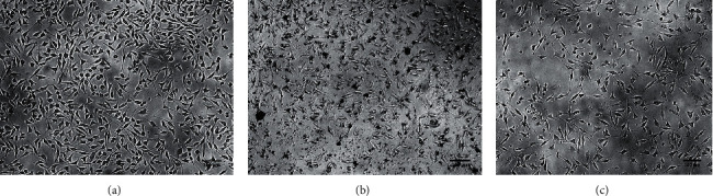
Results: Premna integrifolia extract did not possess any noticeable cytotoxicity to the cell line and showed an IC50 of 185.98 μg/ml. The wound contraction potential of PIE ointment-treated animals was considerably greater (P ≤ 0.001) on days 4, 8, 12, 16, and 20 when compared to the control group. The percentage of wound contraction on day 20 was 99.92% in PIE-treated animals compared to 83.23% in untreated animals. Compared to the untreated group, the duration of full epithelization was significantly (P ≤ 0.01) shorter in the test group. When compared to the incision control group, the animals treated with PIE ointment had significantly higher (P ≤ 0.001) tensile strength. In addition, animals given the test drug had a significant (P ≤ 0.001) increase in total protein and hydroxyproline. In the in vitro scratch assay, test drug-treated cells demonstrated greater cell migration. Histology images confirmed that the test drug-treated group had epithelial tissue proliferation and keratinization.
Conclusion: The current study found that Premna integrifolia improved wound healing activity both in vitro and in vivo. These findings indicate that Premna integrifolia extract has wound-healing potential and could be a viable source of nutraceuticals with wound-healing properties.
Copyright © 2022 Saeed Ali Alsareii et al.
Conflict of interest statement
The authors declare no conflicts of interest.
Figures


References
-
- Mulkalwar S., Behera L., Golande P., Manjare R., Patil H. Evaluation of wound healing activity of topical phenytoin in an excision wound model in rats. International Journal of Basic & Clinical Pharmacology . 2015;4(1):p. 139. doi: 10.5455/2319-2003.ijbcp20150225. - DOI
-
- Shrimanker M., Patel N., Modi H., Dave R. A review: screening models for wound healing activity in animals. American Journal of PharmTech Research . 2013;3:2249–3387.
LinkOut - more resources
Full Text Sources
