

Localization of the Center of the Intramuscular Nerve Dense Region of the Suboccipital Muscles: An Anatomical Study
- PMID: 35463128
- PMCID: PMC9019081
- DOI: 10.3389/fneur.2022.863446
Localization of the Center of the Intramuscular Nerve Dense Region of the Suboccipital Muscles: An Anatomical Study
Abstract
Purpose: This study aimed to determine the body surface puncture position and depth of the center of the intramuscular nerve dense region in the suboccipital muscle to provide morphological guidance for accurate botulinum toxin A injection to treat headaches caused by increased suboccipital muscle tension.
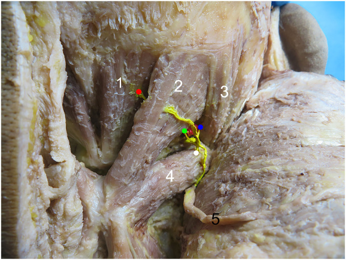
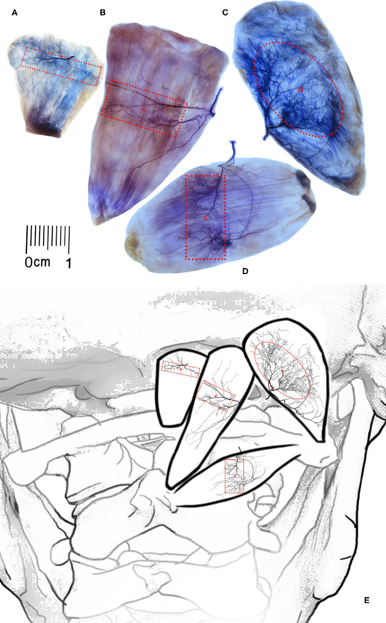
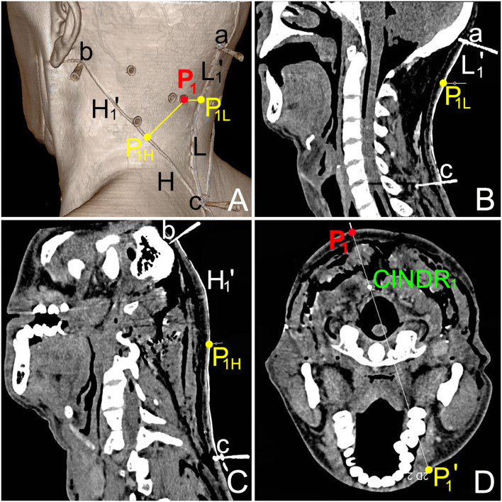
Methods: Twenty-four cadavers aged 66.5 ± 5.3 years were studied. The curve line connecting occipital eminence or mastoid process and spinous process of the 7th cervical vertebrae was considered the longitudinal reference line (L) and horizontal reference line (H), respectively. Sihler's staining, barium sulfate labeling, and CT were employed. The body surface projection point of the center of the intramuscular nerve dense region was designated as P. The projection of the center of the intramuscular nerve dense region was in the opposite direction across the transverse plane and was recorded as P'. The intersections of the vertical line through point P and lines L and H were designated as PL and PH. The percentage position of the PH and PL points on the H and L lines and the depths of the center of intramuscular nerve dense regions were identified.
Results: Sihler's staining showed one intramuscular nerve-dense region in each suboccipital muscle. The PH of the center of the intramuscular nerve dense region was located at 51.40, 45.55, 20.55, and 43.50%. The PL was located at 31.38, 30.08, 16.91, and 52.94%. The depth of the center of the intramuscular nerve dense region was at 22.26, 22.54, 13.14, and 27.30%. These percentage values are all the means.
Conclusion: Accurately defining the body surface position and depth of the center of intramuscular nerve dense region in suboccipital muscles will help to improve botulinum toxin A to target localization efficiency for treating tension-type headache.
Keywords: botulinum toxin A; center of intramuscular nerve dense region; suboccipital muscle; target localization; tension-type headaches.
Copyright © 2022 Wang, Li, Wang and Yang.
Conflict of interest statement
The authors declare that the research was conducted in the absence of any commercial or financial relationships that could be construed as a potential conflict of interest.
Figures
Similar articles
-
Division of neuromuscular compartments and localization of the center of the intramuscular nerve-dense region in pelvic wall muscles based on Sihler's staining.Anat Sci Int. 2024 Jan;99(1):127-137. doi: 10.1007/s12565-023-00744-4. Epub 2023 Sep 28. Anat Sci Int. 2024. PMID: 37768515 Free PMC article.
-
Localization of nerve entry points and the center of intramuscular nerve-dense regions in the adult pectoralis major and pectoralis minor and its significance in blocking muscle spasticity.J Anat. 2021 Nov;239(5):1123-1133. doi: 10.1111/joa.13493. Epub 2021 Jun 27. J Anat. 2021. PMID: 34176122 Free PMC article.
-
Localization of the center of the intramuscular nerve dense region of the medial femoral muscles and the significance for blocking spasticity.Ann Anat. 2020 Sep;231:151529. doi: 10.1016/j.aanat.2020.151529. Epub 2020 May 11. Ann Anat. 2020. PMID: 32437866
-
Sihler's staining technique: How to and guidance for botulinum neurotoxin injection in human muscles.Clin Anat. 2024 Mar;37(2):169-177. doi: 10.1002/ca.24076. Epub 2023 May 31. Clin Anat. 2024. PMID: 37255275 Review.
-
Innervation of the Face Studied Using Modifications to Sihler's Technique in a Primate Model.Plast Reconstr Surg. 2008 Apr;121(4):1188-1205. doi: 10.1097/01.prs.0000305563.77782.35. Plast Reconstr Surg. 2008. PMID: 18349636 Review.
Cited by
-
Localisation of the centre of the highest region of muscle spindle abundance of anterior forearm muscles.J Anat. 2024 May;244(5):803-814. doi: 10.1111/joa.14000. Epub 2023 Dec 28. J Anat. 2024. PMID: 38155435 Free PMC article.
-
The highest region of muscle spindle abundance should be the optimal target of botulinum toxin A injection to block muscle spasms in rats.Front Neurol. 2023 Feb 23;14:1061849. doi: 10.3389/fneur.2023.1061849. eCollection 2023. Front Neurol. 2023. PMID: 36908586 Free PMC article.
-
Division of neuromuscular compartments and localization of the center of the intramuscular nerve-dense region in pelvic wall muscles based on Sihler's staining.Anat Sci Int. 2024 Jan;99(1):127-137. doi: 10.1007/s12565-023-00744-4. Epub 2023 Sep 28. Anat Sci Int. 2024. PMID: 37768515 Free PMC article.
References
LinkOut - more resources
Full Text Sources