Erdheim-Chester Disease Revealed by Central Positional Nystagmus: A Case Report
- PMID: 35463141
- PMCID: PMC9022006
- DOI: 10.3389/fneur.2022.880312
Erdheim-Chester Disease Revealed by Central Positional Nystagmus: A Case Report
Abstract
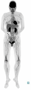
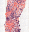
Erdheim-Chester disease (ECD) is a rare histiocytic disorder, recently recognized to be neoplastic. The clinical phenotype of the disease is extremely heterogeneous, and depends on the affected organs, with the most frequently reported manifestations being bone pain, diabetes insipidus and neurological disorders including ataxia. In this article, we report on a case of a 48-year-old woman, whose initial symptom of gait instability was isolated. This was associated with positional nystagmus with central features: nystagmus occurring without latency, clinically present with only mild symptoms, and resistant to repositioning maneuvers. The cerebral MRI showed bilateral intra-orbital retro-ocular mass lesions surrounding the optic nerves and T2 hyperintensities in the pons and middle cerebellar peduncles. A subsequent CT scan of the chest abdomen and pelvis found a left "hairy kidney", while 18 F-FDG PET-CT imaging disclosed symmetric 18F-FDG avidity predominant at the diametaphyseal half of both femurs. Percutaneous US-guided biopsy of perinephric infiltrates and the kidney showed infiltration by CD68(+), CD1a(-), Langerin(-), PS100(-) foamy histiocytes with BRAF V600E mutation. The combination of the different radiological abnormalities and the result of the biopsy confirmed the diagnosis of ECD. Many clinical and radiological descriptions are available in the literature, but few authors describe vestibulo-ocular abnormalities in patients with ECD. Here, we report on a case of ECD and provide a precise description of the instability related to central positional nystagmus, which led to the diagnosis of ECD.
Keywords: Erdheim-Chester disease; case report; central positional nystagmus; dizziness; gait; nystagmus.
Copyright © 2022 Weckel, Gallois, Debs, Escude, Tremelet, Varenne, Biotti, Chauveau and Bonneville.
Conflict of interest statement
The authors declare that the research was conducted in the absence of any commercial or financial relationships that could be construed as a potential conflict of interest.
Figures


References
Publication types
LinkOut - more resources
Full Text Sources
Research Materials

