Review
. 2022 Apr 7:16:866999.
doi: 10.3389/fncir.2022.866999. eCollection 2022.

The Role of Inhibitory Interneurons in Circuit Assembly and Refinement Across Sensory Cortices

Affiliations
Review

The Role of Inhibitory Interneurons in Circuit Assembly and Refinement Across Sensory Cortices

Camilo Ferrer et al. Front Neural Circuits. .

Abstract

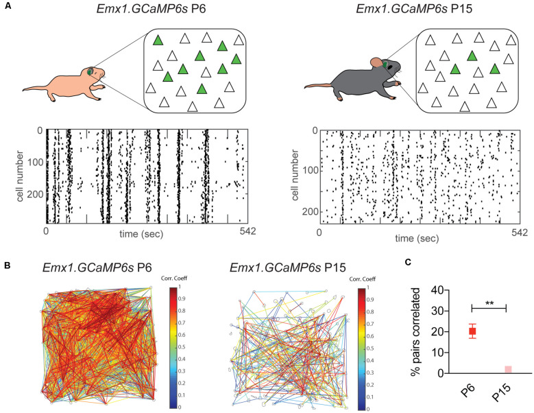
Sensory information is transduced into electrical signals in the periphery by specialized sensory organs, which relay this information to the thalamus and subsequently to cortical primary sensory areas. In the cortex, microcircuits constituted by interconnected pyramidal cells and inhibitory interneurons, distributed throughout the cortical column, form the basic processing units of sensory information underlying sensation. In the mouse, these circuits mature shortly after birth. In the first postnatal week cortical activity is characterized by highly synchronized spontaneous activity. While by the second postnatal week, spontaneous activity desynchronizes and sensory influx increases drastically upon eye opening, as well as with the onset of hearing and active whisking. This influx of sensory stimuli is fundamental for the maturation of functional properties and connectivity in neurons allocated to sensory cortices. In the subsequent developmental period, spanning the first five postnatal weeks, sensory circuits are malleable in response to sensory stimulation in the so-called critical periods. During these critical periods, which vary in timing and duration across sensory areas, perturbations in sensory experience can alter cortical connectivity, leading to long-lasting modifications in sensory processing. The recent advent of intersectional genetics, in vivo calcium imaging and single cell transcriptomics has aided the identification of circuit components in emergent networks. Multiple studies in recent years have sought a better understanding of how genetically-defined neuronal subtypes regulate circuit plasticity and maturation during development. In this review, we discuss the current literature focused on postnatal development and critical periods in the primary auditory (A1), visual (V1), and somatosensory (S1) cortices. We compare the developmental trajectory among the three sensory areas with a particular emphasis on interneuron function and the role of inhibitory circuits in cortical development and function.

Keywords: circuit; cortex; critical period; development; inhibition; interneuron; plasticity; sensory.


Conflict of interest statement

The authors declare that the research was conducted in the absence of any commercial or financial relationships that could be construed as a potential conflict of interest.

Figures

Figure 1
Figure 1
Timeline comparison of cortical activity development in A1, V1, and S1. Development of cortical activity in mice from P0 to P40 in A1, V1, and S1. In the three areas, synchronized spontaneous activity is present during the first postnatal week, but it desynchronizes through the second postnatal week in A1 (Babola et al., ; Meng et al., 2020); V1 (Rochefort et al., ; Ackman et al., ; Siegel et al., 2012), and S1 (Golshani et al., ; Che et al., 2018). In A1 (top panel), the onset of evoked sensory activity takes place by the end of the first postnatal week and it becomes more prominent upon ear opening at P11 (Anthwal and Thompson, ; Makarov et al., 2021). Two auditory critical periods for pure tones (PT CP; Barkat et al., 2011) and Frequency modulated sweeps (FMS CP; Bhumika et al., 2020) take place shortly after, P12–15 and P31–38 respectively. In V1 (middle panel), evoked visual responses start by the end of the first postnatal week and become more reliable by the second postnatal week (Colonnese et al., 2010), around the time of eye opening (~P14). The critical period for ocular dominance (ODP CP) takes place between P21 and P35 (Hensch, ; Espinosa and Stryker, 2012) and an orientation selectivity (OS) CP has been shown in google-reared mice between P28 and P49 (Yoshida et al., 2012). In S1 (bottom panel), passive whisker stimulation can trigger evoked responses from birth (Anton-Bolanos et al., 2019), but these responses become more reliable around P6–8 (Colonnese et al., 2010) and active whisking onset occurs around P14 (Landers and Philip Zeigler, 2006). An early anatomical CP is observed in S1 from P0 to P4, capable of altering barrel formation and thalamocortical innervation (Durham and Woolsey, ; Lee et al., 2009). While throughout life, different paradigms of sensory deprivation can induce discrete forms experience-dependent plasticity in particular layers/synapses (Nowicka et al., ; Wen and Barth, ; Gainey et al., 2018).
Figure 2
Figure 2
Sparsification of spontaneous cortical activity in S1 by the end of the second postnatal week. Desynchronization of spontaneous cortical activity in S1 measured using in vivo calcium imaging. (A) Top panel: diagrams of in vivo imaging of Emx1.GCaMP6 s mice at P6 and P15, with cranial windows placed over S1. The number of spontaneously co-active Pyr neurons (green) at any given time decreases between P6 and P15. Bottom panel: representative raster plots of neuronal calcium activity at both ages. Each row represents a single neuron imaged during 542 s. Each tick represents the onset of a single calcium event. At P6 neuronal activity is characterized by the co-activation of most neurons imaged in the field of view, visualized in vertical arrangements in the raster plot, while at P15 neuronal activity becomes less synchronized resulting in a “salt and pepper” pattern in the raster plot. (B) Visualization of correlated neuronal activity corresponding to recordings in (A). Gray contours indicate detected somas in which calcium signals were analyzed. Significantly correlated cell pairs are connected by lines. Line color indicates the magnitude of the correlation coefficient of the connecting pair. (C) Percentage of pairs that are significantly correlated decreases from P6 to P15. Unpaired t-test, **p = 0.0027. Derived with permission from Che et al. (2018).
Figure 3
Figure 3
Molecular and synaptic loci of plasticity during sensory critical periods. Cortical circuit schematic with major synaptic connections and molecular factors regulating critical period plasticity. The circuit depicted shows the circuit motifs and cellular components regulating CP plasticity: basket PV INs (Brown) inhibit one another and exert strong inhibitory control of Pyr (light blue) through perisomatic inhibition; layer I INs (dark blue) inhibit the apical dendrites of Pyr and can also target PV cells, resulting in simultaneous Pyr somatic disinhibition and dendritic inhibition; Martinoti SST INs (yellow) across layers target Pyr apical dendrites and also receive input from VIP INs (pink), such that VIP activation results in Pyr disinhibition; while a subset layer IV SST INs target PV INs preferentially, resulting in disinhibition; and astrocytes regulate the extracellular matrix and PNN formation onto PV INs. PV IN maturation determines both the onset and closure of cortical critical periods. Maturation of PV intrinsic properties, synaptic inputs (both excitatory and inhibitory, dashed circles), and inhibitory synaptic output onto Pyr (dashed circle) are all crucial for CP plasticity. Expression of KV3.1, GAD65 and NRG1/ErbB4 in PV INs promote (green) normal CP plasticity (Hensch et al., ; Matsuda et al., ; Zheng et al., 2021), while PNN (brown shadow) maturation in PV INs prevent and close CP plasticity (Pizzorusso et al., ; Nowicka et al., ; Sigal et al., 2019). In addition, GABAAα1 receptor expression in Pyr (in putative PV synapses) is necessary for CP plasticity (Fagiolini et al., 2004). Both SST and LI INs can induce CP plasticity indirectly by means of PV IN inhibition, such that the expression of molecular factors promoting SST (Lypd6 and nAChRα2) or LI IN (Lynx1 and nAChRs) activity enhance plasticity (Takesian et al., ; Sadahiro et al., 2020). On the other hand, VIP IN-mediated Pyr disinhibition, via SST inhibition, also promotes cortical plasticity (Fu et al., 2015). In contrast, connexin 30 expression in astrocytes restricts CP plasticity via PNN maturation in PV INs (Ribot et al., 2021). Green font/arrows represent molecules or synapses promoting CP plasticity, while red font/arrows represent those preventing plasticity. Abbreviations: PV, Parvalbumin; Pyr, Pyramidal cell; SST, Somatostatin; LI INs, Layer I interneurons; VIP, Vasoactive intestinal peptide; PNN, Perineuronal net; nAChRs, Nicotinic Acetylcholine receptors; NRG1, Neuregulin 1; KV3.1, Potassium channel 3.1; GAD65, Glutamic acid decarboxylase 65-kilodalton.

Similar articles

Cited by

References

    1. Abs E., Poorthuis R. B., Apelblat D., Muhammad K., Pardi M. B., Enke L., et al. . (2018). Learning-Related Plasticity in Dendrite-Targeting Layer 1 Interneurons. Neuron 100, 684–699.e6. 10.1016/j.neuron.2018.09.001 - DOI - PMC - PubMed
    1. Ackman J. B., Burbridge T. J., Crair M. C. (2012). Retinal waves coordinate patterned activity throughout the developing visual system. Nature 490, 219–225. 10.1038/nature11529 - DOI - PMC - PubMed
    1. Adesnik H., Bruns W., Taniguchi H., Huang Z. J., Scanziani M. (2012). A neural circuit for spatial summation in visual cortex. Nature 490, 226–231. 10.1038/nature11526 - DOI - PMC - PubMed
    1. Agmon A., Yang L. T., O’Dowd D. K., Jones E. G. (1993). Organized growth of thalamocortical axons from the deep tier of terminations into layer IV of developing mouse barrel cortex. J. Neurosci. 13, 5365–5382. 10.1523/JNEUROSCI.13-12-05365.1993 - DOI - PMC - PubMed
    1. Akgul G., Wollmuth L. P. (2013). Synapse-associated protein 97 regulates the membrane properties of fast-spiking parvalbumin interneurons in the visual cortex. J. Neurosci. 33, 12739–12750. 10.1523/JNEUROSCI.0040-13.2013 - DOI - PMC - PubMed

Publication types

LinkOut - more resources