

Published Erratum
doi: 10.3389/fonc.2022.793448.
eCollection 2022.
Corrigendum: Downregulation of GLYAT Facilitates Tumor Growth and Metastasis and Poor Clinical Outcomes Through the PI3K/AKT/Snail Pathway in Human Breast Cancer
Affiliations
- PMID: 35463385
- PMCID: PMC9020255
- DOI: 10.3389/fonc.2022.793448
Item in Clipboard
Published Erratum
Corrigendum: Downregulation of GLYAT Facilitates Tumor Growth and Metastasis and Poor Clinical Outcomes Through the PI3K/AKT/Snail Pathway in Human Breast Cancer
Front Oncol.
.
Abstract
[This corrects the article DOI: 10.3389/fonc.2021.641399.].
Keywords: EMT; GLYAT; PI3K/AKT; breast cancer; clinicopathological features; prognosis.
Copyright © 2022 Tian, Wu, Jiang, Zhang, Wu, Miao, Liu and Gao.
Figures

GLYAT suppresses BC cell proliferation and metastasis. (A) GLYAT protein level was assessed in different BC cell lines. (B) MDA-MB-231 cells were transfected with the GLYAT KD plasmid and a scrambled plasmid as control. The suppression of GLYAT in MDA-MB-231 cells was confirmed at the protein level. (C) MCF-7 cells were transfected with the GLYAT OE plasmid and a scrambled plasmid as control. The overexpression of GLYAT in MCF-7 cells was confirmed at the protein level. (D, E) GLYAT KD in MDA-MB-231 cells significantly increased colony numbers and size compared with the scramble cells. (F) The Transwell assay revealed that the migration ability of MDA-MB-231 cells was significantly increased following transfection with GLYAT KD1 and KD2 compared with the scramble control. (G) The Transwell assay revealed that the migratory ability was significantly decreased in GLYAT OE MCF-7 cells compared with scramble control. **p < 0.01. Scale bars for (E) are 100 μm and 25 μm, and for (F, G) are 100 μm.
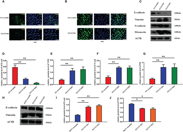
GLYAT suppresses EMT phenotype in BC cells. (A, B) The immunofluorescence assay showed that E-cadherin expression was decreased and vimentin expression was increased after the treatment of GLYAT KD. (C–G) Protein levels of E-cadherin was reduced, whereas the levels of vimentin, N-cadherin, and fibronectin were increased in MDA-MB-231 cells following GLYAT inhibition. (H–J) Protein levels of E-cadherin were increased whereas the expression of vimentin was reduced in GLYAT OE MCF-7 cells. *p < 0.05, **p < 0.01. Scale bars are 25 μm.
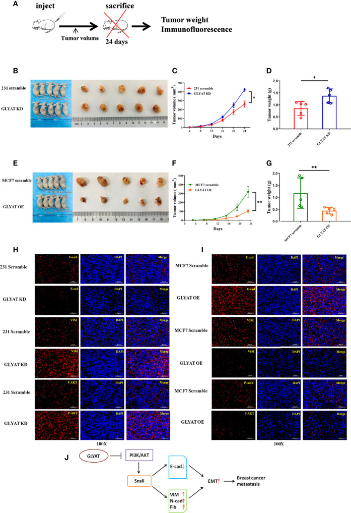
GLYAT suppresses breast cancer proliferation, metastasis, and EMT in vivo. (A) Schematic diagram of the metastasis model in mice. (B–D) The mice injected with stable GLYAT KD MDA-MB-231 cells had markedly bigger and heavier tumors. (E–G) The mice injected with stable GLYAT OE MCF-7 cells had markedly smaller and lighter tumors. (H) The immunofluorescence assay of serial sections of mouse tumor tissues revealed that GLYAT KD MDA-MB-231 cells significantly decreased the expression of E-cadherin whereas increased the expression of vimentin and p-AKT. (I) The immunofluorescence assay of serial sections of mouse tumor tissues revealed that, in GLYAT OE MCF-7 cells, the expression of E-cadherin was increased whereas vimentin and p-AKT were decreased. (J) The schematic diagram of the role of GLYAT in BC. *p < 0.05, **p < 0.01. Scale bars are 50 mm.
Erratum for
-
Downregulation of GLYAT Facilitates Tumor Growth and Metastasis and Poor Clinical Outcomes Through the PI3K/AKT/Snail Pathway in Human Breast Cancer.Front Oncol. 2021 Apr 22;11:641399. doi: 10.3389/fonc.2021.641399. eCollection 2021. Front Oncol. 2021. PMID: 33968740 Free PMC article.
Publication types
LinkOut - more resources
Full Text Sources
Research Materials