. 2022 Apr;38(Suppl 1):91-100.
doi: 10.1007/s12055-020-01125-6. Epub 2021 Feb 22.

Aortic root widening: "pro et contra"

Affiliations

Aortic root widening: "pro et contra"

Balaji Srimurugan et al. Indian J Thorac Cardiovasc Surg. 2022 Apr.

Abstract

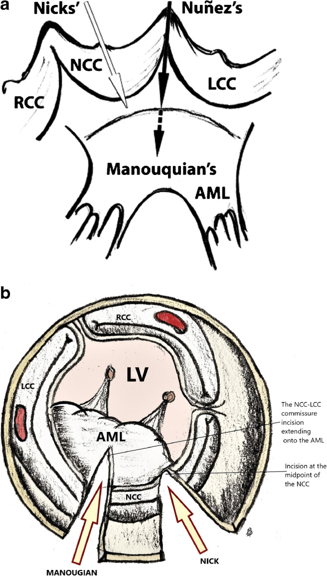
In patients with a small aortic annulus, the clinical benefits of aortic valve replacement depend on avoidance of patient-prosthesis mismatch as it is associated with reduced overall survival. Aortic root widening or enlargement is a useful technique to implant larger valve prosthesis to prevent patient-prosthesis mismatch. Posterior annular enlargement is the commonest technique used for aortic root enlargement. Consistent enlargement of the aortic root requires more extensive procedures like Manouguian or Konno-Rastan techniques. The patients commonly selected are younger patients with good life expectancy. However, caution is advised in applying this procedure in elderly patients, patients with heavily calcified annulus and when performing concomitant procedures. There is no definitive conclusion on the best material to use for the reconstruction of aortic annulus and aorta in aortic root enlargement procedures.

Keywords: Aortic root enlargement; Aortic valve stenosis; Manouguian; Nicks; Patient-prosthesis mismatch.


Conflict of interest statement

Conflict of interestThe authors declare that they have no conflicts of interest.

Figures

Fig. 1
Fig. 1
a, b Aortic root anatomy—reproduced with permission from reference . True ventricular- aortic junction is above the basal ring. Crown-shaped valves form the hemodynamic junction. When they are excised, as for AVR, the remnants form the surgical annulus, which is also crown-shaped
Fig. 2
Fig. 2
The valves are sized at the basal ring, while the sutures are placed at the crown shaped surgical annulus. Valve sizers are based on tissue annulus diameter. In intra-annular implantation, part of sewing ring also needs to be accommodated into the annulus which can result in valve not properly “fitting” in to the annulus. This can be avoided by supra-annular placement. Supra-annular valves run the risk of coronary ostia obstruction, especially in bicuspid valves, where para-commissural origin of coronaries is not uncommon. Since the sino-tubular junction is smaller than annulus, there can be considerable difficulty in lowering the valve to the annulus and for tying the sutures
Fig. 3
Fig. 3
a, b The commonly performed posterior enlargement techniques. Antunes technique is similar to Nicks; however, it extends below the aortic annular ring to aorto-mitral curtain. a Reproduced with permission from reference [34]. RCC, right coronary cusp; NCC, non-coronary cusp; LCC, left coronary cusp; AML, anterior mitral leaflet; LV, left ventricle
Fig. 4
Fig. 4
a–c Steps in Nick’s procedure. The patch material can be pericardium (autologous or bovine) or prosthetic material like Dacron or coated graft. Sometimes, upsizing requires tilted positioning of the valve. RCC, right coronary cusp; NCC, non-coronary cusp; LCC, left coronary cusp; AML, anterior mitral leaflet; LV, left ventricle
Fig. 5
Fig. 5
a–c Steps in the Manouguian procedure. It is essential to make the incision on the center of anterior mitral leaflet in the clear zone. The center of anterior mitral leaflet does not necessarily correspond to the left and non-coronary commissure. Otherwise, it can induce mitral regurgitation. RCC, right coronary cusp; NCC, non-coronary cusp; LCC, left coronary cusp; AML, anterior mitral leaflet; LV, left ventricle; LA, left atrium
Fig. 6
Fig. 6
a–c Steps of the Konno–Rastan procedure. The aortotomy incision is extended to inter-ventricular septum, well leftward from the right coronary orifice. The inter-ventricular septal incision runs the risk of injury to first septal artery. RV, right ventricle; LV, left ventricle; IVS, inter-ventricular septum; RA, right atrium; Ao, aorta; PA, pulmonary artery
Fig. 7
Fig. 7
a, b. Right ventricular outflow patch is anchored to the left ventricular patch. The procedure is often talked about, but seldom performed. RVOT, right ventricle outflow tract; LVOT, left ventricle outflow tract; IVS, inter-ventricular septum; RA, right atrium; Ao, aorta; PA, pulmonary artery

References

    1. Horstkotte D, Loogen F. The natural history of aortic valve stenosis. Eur Heart J. 1988;9:57–64. - PubMed
    1. Nishimura RA, Otto CM, Bonow RO, et al. 2014 AHA/ACC guideline for the management of patients with valvular heart disease: executive summary: a report of the American College of Cardiology/American Heart Association Task Force on Practice Guidelines. Circulation. 2014;129:2440–2492. - PubMed
    1. Chambers J. Echocardiography and the small aortic root. J Heart Valve Dis. 1996;5:S264–S268. - PubMed
    1. Ghosh P, Kumar S, Pandey S, Kumar AS, Sinha N. Small aortic annulus: a functional definition. Ann Thorac Cardiovasc Surg. 1998;4:251–261. - PubMed
    1. Freitas-Ferraz AB, Tirado-Conte G, Dagenais F, et al. Aortic stenosis and small aortic annulus. Circulation. 2019;139:2685–2702. - PubMed

LinkOut - more resources