

Abdominal Aortic Endothelial Dysfunction Occurs in Female Mice With Dextran Sodium Sulfate-Induced Chronic Colitis Independently of Reactive Oxygen Species Formation
- PMID: 35463755
- PMCID: PMC9021429
- DOI: 10.3389/fcvm.2022.871335
Abdominal Aortic Endothelial Dysfunction Occurs in Female Mice With Dextran Sodium Sulfate-Induced Chronic Colitis Independently of Reactive Oxygen Species Formation
Abstract
Background and objective: Inflammatory bowel disease (IBD) produces significant local and systemic inflammation with increased reactive oxygen species (ROS) formation. IBD Patients are at an increased risk for developing endothelial dysfunction and cardiovascular diseases. The present study tested the hypothesis that IBD impairs aortic endothelial function via ROS formation and investigate potential sex-related differences.
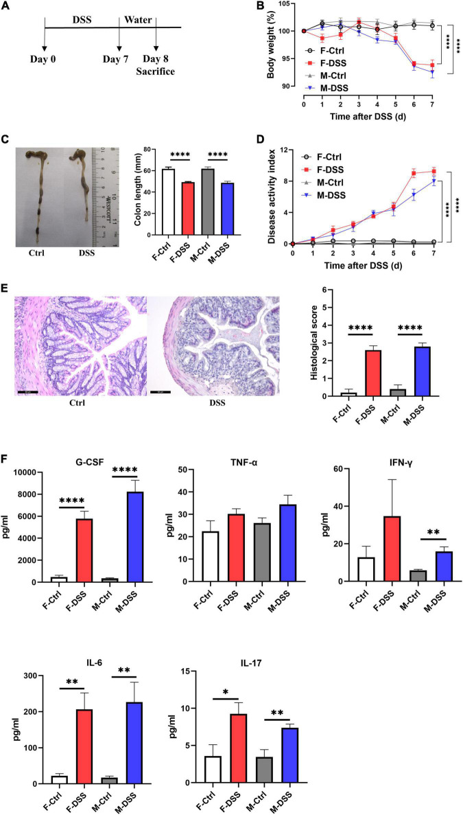
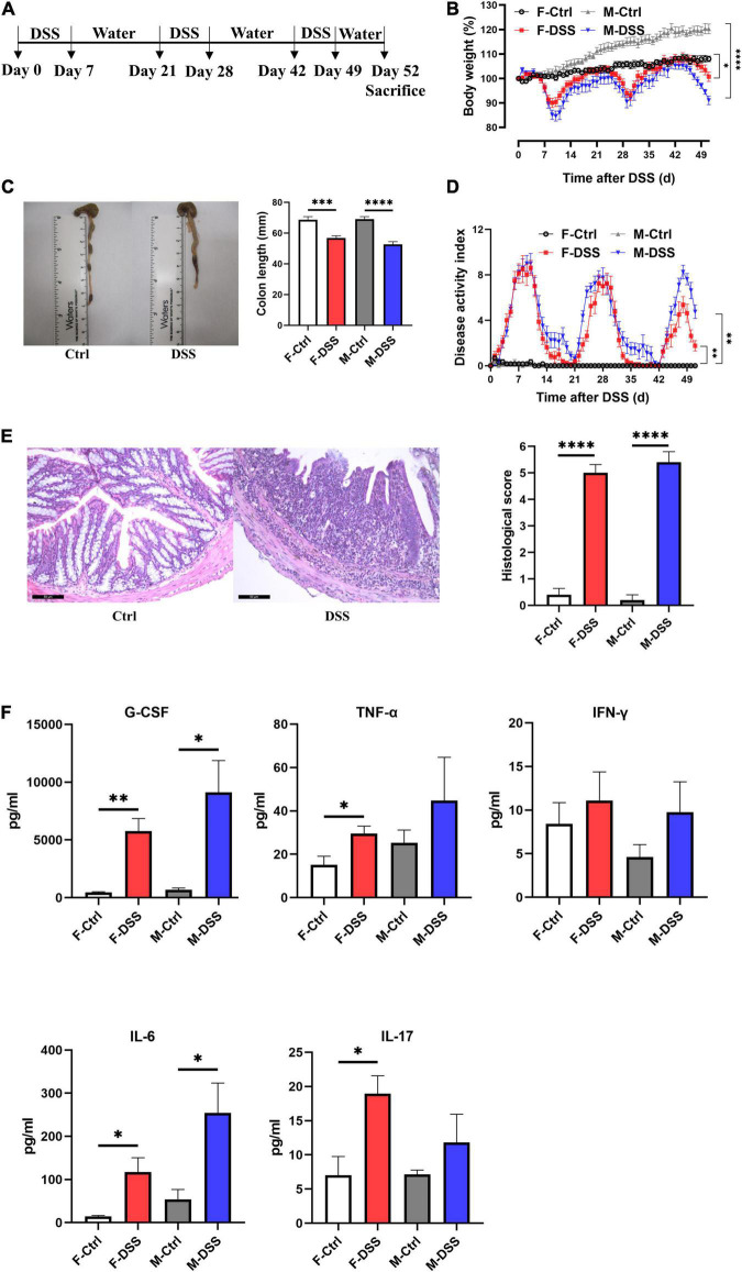
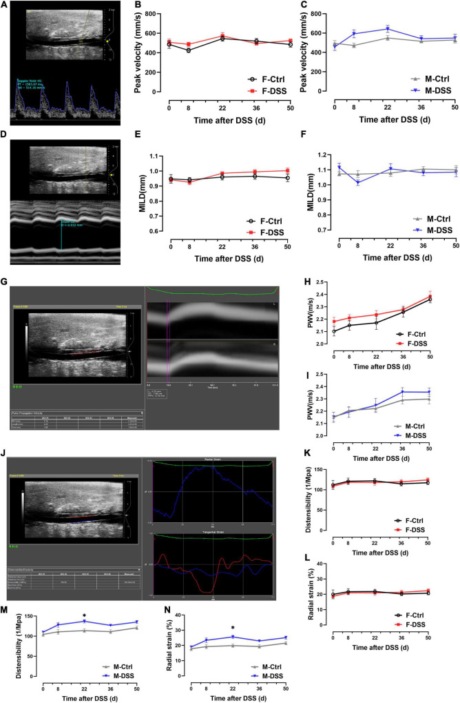
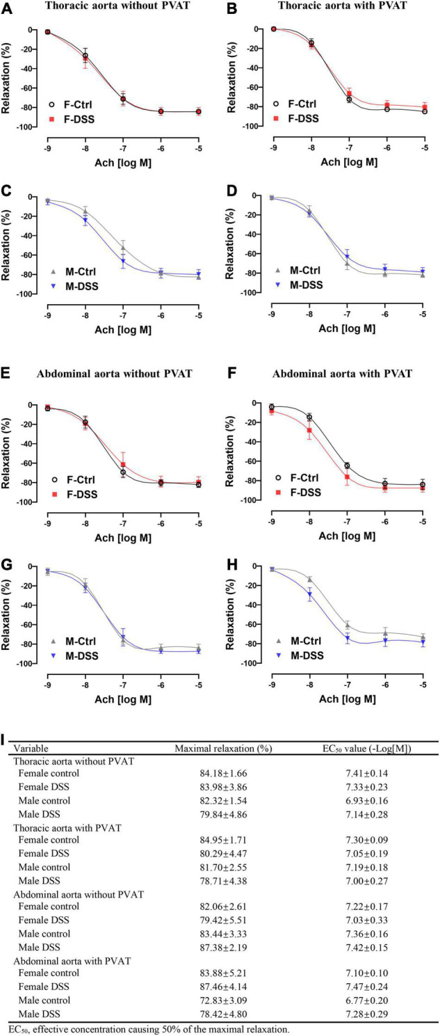
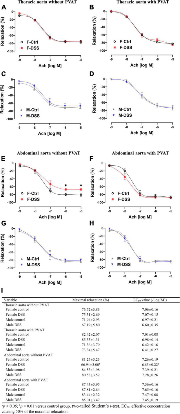
Methods and results: Acute and chronic colitis models were induced in male and female C57BL/6 mice with dextran sodium sulfate (DSS) treatment. Aortic wall stiffness, endothelial function, and ROS levels, as well as serum levels of pro-inflammatory cytokines were evaluated. Acetylcholine (Ach)-induced endothelium-dependent relaxation of abdominal aorta without perivascular adipose tissue (PVAT) was significantly reduced in female mice, not males, with chronic colitis without a change in nitroglycerin-induced endothelium-independent relaxation. PVAT effectively preserved Ach-induced relaxation in abdominal aorta of female mice with chronic colitis. Aortic peak velocity, maximal intraluminal diameters, pulse wave velocity, distensibility and radial strain were preserved in mice with both acute and chronic colitis. Although pro-inflammatory cytokines levels were increased in mice with acute and chronic colitis, aortic ROS levels were not increased.
Conclusion: The data demonstrate that abdominal aortic endothelial function was attenuated selectively in female mice with chronic colitis independent of ROS formation. Further, PVAT played an important role in preserving endothelial function in female mice with chronic colitis.
Keywords: ROS; aorta; colitis; endothelial dysfunction; inflammatory bowel disease.
Copyright © 2022 Wu, Hu, Zhang, Xia, Liu, Zhu, Wang, Sun, Hao, Cui, Parrish, Li, Hill, Xu and Liu.
Conflict of interest statement
The authors declare that the research was conducted in the absence of any commercial or financial relationships that could be construed as a potential conflict of interest.
Figures

Similar articles
-
Recovery of Ischemic Limb and Femoral Artery Endothelial Function Are Preserved in Mice with Dextran Sodium Sulfate-Induced Chronic Colitis.Biology (Basel). 2022 Aug 4;11(8):1169. doi: 10.3390/biology11081169. Biology (Basel). 2022. PMID: 36009796 Free PMC article.
-
Gestational intermittent hypoxia induces endothelial dysfunction, reduces perivascular adiponectin and causes epigenetic changes in adult male offspring.J Physiol. 2019 Nov;597(22):5349-5364. doi: 10.1113/JP277936. Epub 2019 Aug 22. J Physiol. 2019. PMID: 31441069
-
Helicobacter pylori infection selectively attenuates endothelial function in male mice via exosomes-mediated ROS production.Front Cell Infect Microbiol. 2023 May 18;13:1142387. doi: 10.3389/fcimb.2023.1142387. eCollection 2023. Front Cell Infect Microbiol. 2023. PMID: 37274312 Free PMC article.
-
Prdx6 Deficiency Ameliorates DSS Colitis: Relevance of Compensatory Antioxidant Mechanisms.J Crohns Colitis. 2017 Jul 1;11(7):871-884. doi: 10.1093/ecco-jcc/jjx016. J Crohns Colitis. 2017. PMID: 28199527
-
Different Anti-Contractile Function and Nitric Oxide Production of Thoracic and Abdominal Perivascular Adipose Tissues.Front Physiol. 2016 Jul 12;7:295. doi: 10.3389/fphys.2016.00295. eCollection 2016. Front Physiol. 2016. PMID: 27462277 Free PMC article.
Cited by
-
Recovery of Ischemic Limb and Femoral Artery Endothelial Function Are Preserved in Mice with Dextran Sodium Sulfate-Induced Chronic Colitis.Biology (Basel). 2022 Aug 4;11(8):1169. doi: 10.3390/biology11081169. Biology (Basel). 2022. PMID: 36009796 Free PMC article.
-
Adventitial macrophage accumulation impairs perivascular nerve function in mesenteric arteries with inflammatory bowel disease.Front Physiol. 2023 May 25;14:1198066. doi: 10.3389/fphys.2023.1198066. eCollection 2023. Front Physiol. 2023. PMID: 37342800 Free PMC article.
References
Grants and funding
LinkOut - more resources
Full Text Sources