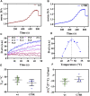
Disease-Related Protein Variants of the Highly Conserved Enzyme PAPSS2 Show Marginal Stability and Aggregation in Cells
- PMID: 35463959
- PMCID: PMC9024126
- DOI: 10.3389/fmolb.2022.860387
Disease-Related Protein Variants of the Highly Conserved Enzyme PAPSS2 Show Marginal Stability and Aggregation in Cells
Abstract
Cellular sulfation pathways rely on the activated sulfate 3'-phosphoadenosine-5'-phosphosulfate (PAPS). In humans, PAPS is exclusively provided by the two PAPS synthases PAPSS1 and PAPSS2. Mutations found in the PAPSS2 gene result in severe disease states such as bone dysplasia, androgen excess and polycystic ovary syndrome. The APS kinase domain of PAPSS2 catalyzes the rate-limiting step in PAPS biosynthesis. In this study, we show that clinically described disease mutations located in the naturally fragile APS kinase domain are associated either with its destabilization and aggregation or its deactivation. Our findings provide novel insights into possible molecular mechanisms that could give rise to disease phenotypes associated with sulfation pathway genes.
Keywords: PAPS synthase; in-cell spectroscopy; protein folding; stability and aggregation; sulfation pathways.
Copyright © 2022 Brylski, Shrestha, House, Gnutt, Mueller and Ebbinghaus.
Conflict of interest statement
The authors declare that the research was conducted in the absence of any commercial or financial relationships that could be construed as a potential conflict of interest.
Figures


Similar articles
-
Human DHEA sulfation requires direct interaction between PAPS synthase 2 and DHEA sulfotransferase SULT2A1.J Biol Chem. 2018 Jun 22;293(25):9724-9735. doi: 10.1074/jbc.RA118.002248. Epub 2018 May 9. J Biol Chem. 2018. PMID: 29743239 Free PMC article.
-
Human 3'-phosphoadenosine 5'-phosphosulfate (PAPS) synthase: biochemistry, molecular biology and genetic deficiency.IUBMB Life. 2003 Jan;55(1):1-11. doi: 10.1080/1521654031000072148. IUBMB Life. 2003. PMID: 12716056 Review.
-
Melting Down Protein Stability: PAPS Synthase 2 in Patients and in a Cellular Environment.Front Mol Biosci. 2019 May 3;6:31. doi: 10.3389/fmolb.2019.00031. eCollection 2019. Front Mol Biosci. 2019. PMID: 31131283 Free PMC article. Review.
-
3'-Phosphoadenosine 5'-phosphosulfate (PAPS) synthases, naturally fragile enzymes specifically stabilized by nucleotide binding.J Biol Chem. 2012 May 18;287(21):17645-17655. doi: 10.1074/jbc.M111.325498. Epub 2012 Mar 26. J Biol Chem. 2012. PMID: 22451673 Free PMC article.
-
Structural basis for the substrate recognition mechanism of ATP-sulfurylase domain of human PAPS synthase 2.Biochem Biophys Res Commun. 2022 Jan 1;586:1-7. doi: 10.1016/j.bbrc.2021.11.062. Epub 2021 Nov 19. Biochem Biophys Res Commun. 2022. PMID: 34818583
Cited by
-
Identification of Genetic Variants for Diabetic Retinopathy Risk Applying Exome Sequencing in Extreme Phenotypes.Biomed Res Int. 2024 Jan 13;2024:2052766. doi: 10.1155/2024/2052766. eCollection 2024. Biomed Res Int. 2024. PMID: 38249632 Free PMC article.
-
Adenylyl-Sulfate Kinase (Met14)-Dependent Cysteine and Methionine Biosynthesis Pathways Contribute Distinctively to Pathobiological Processes in Cryptococcus neoformans.Microbiol Spectr. 2023 Jun 15;11(3):e0068523. doi: 10.1128/spectrum.00685-23. Epub 2023 Apr 10. Microbiol Spectr. 2023. PMID: 37036370 Free PMC article.
-
Phase Separation Oppositely Modulates G-quadruplex and i-Motif DNA Folding in the Nuclei of Living Cells.bioRxiv [Preprint]. 2025 Jul 4:2025.06.30.661982. doi: 10.1101/2025.06.30.661982. bioRxiv. 2025. PMID: 40631133 Free PMC article. Preprint.
-
Heat application in live cell imaging.FEBS Open Bio. 2024 Dec;14(12):1940-1954. doi: 10.1002/2211-5463.13912. Epub 2024 Nov 3. FEBS Open Bio. 2024. PMID: 39489617 Free PMC article. Review.
-
Ramping up the Heat: Induction of Systemic and Pulmonary Immune Responses and Metabolic Adaptations in Mice.bioRxiv [Preprint]. 2025 Aug 2:2025.08.01.667768. doi: 10.1101/2025.08.01.667768. bioRxiv. 2025. PMID: 40766621 Free PMC article. Preprint.
References
-
- Ahmad M., Ul Haque M. F., Ahmad W., Abbas H., Haque S., Krakow D., et al. (1998). Distinct, Autosomal Recessive Form of Spondyloepimetaphyseal Dysplasia Segregating in an Inbred Pakistani kindred. Am. J. Med. Genet. 78, 468–473. 10.1002/(sici)1096-8628(19980806)78:5<468:aid-ajmg13>3.0.co;2-d - DOI - PubMed
LinkOut - more resources
Full Text Sources
Miscellaneous

