. 2022 Apr 8:16:844654.
doi: 10.3389/fnana.2022.844654. eCollection 2022.

Immunocytochemical Localization of Enzymes Involved in Dopamine, Serotonin, and Acetylcholine Synthesis in the Optic Neuropils and Neuroendocrine System of Eyestalks of Paralithodes camtschaticus

Affiliations

Immunocytochemical Localization of Enzymes Involved in Dopamine, Serotonin, and Acetylcholine Synthesis in the Optic Neuropils and Neuroendocrine System of Eyestalks of Paralithodes camtschaticus

Elena Kotsyuba et al. Front Neuroanat. .

Abstract

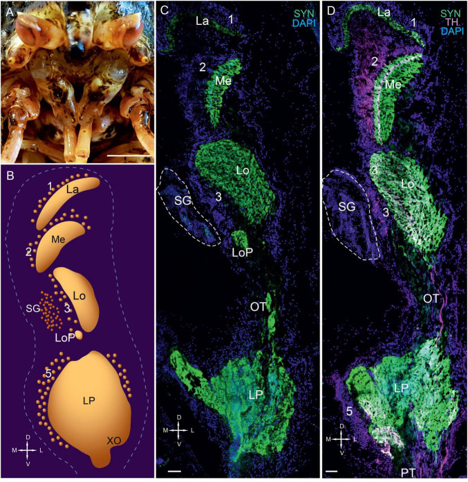
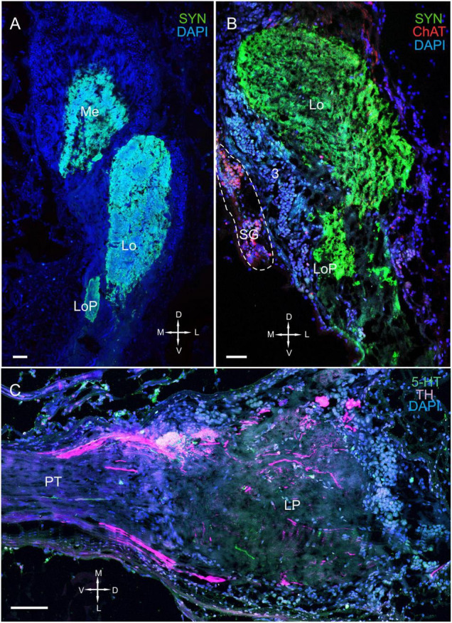
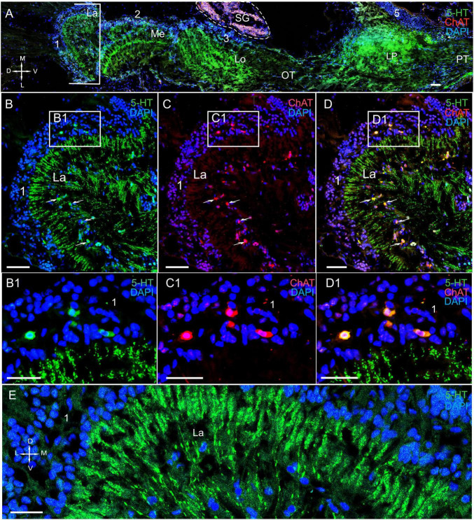
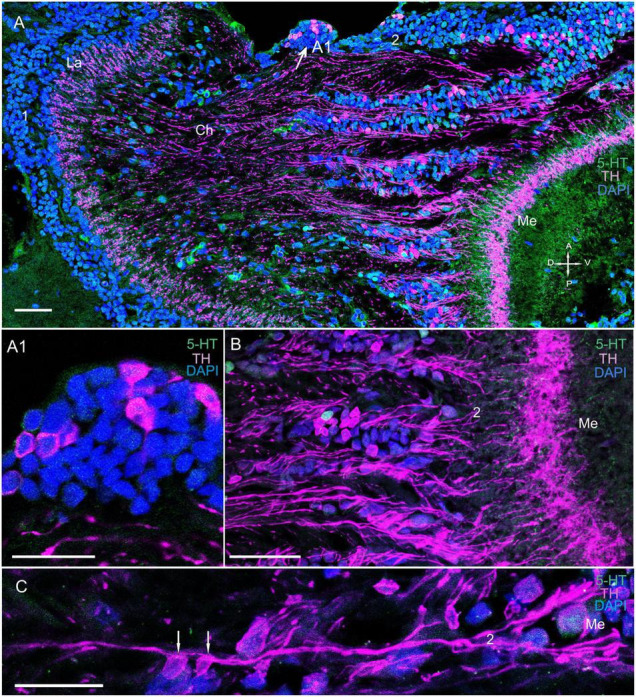
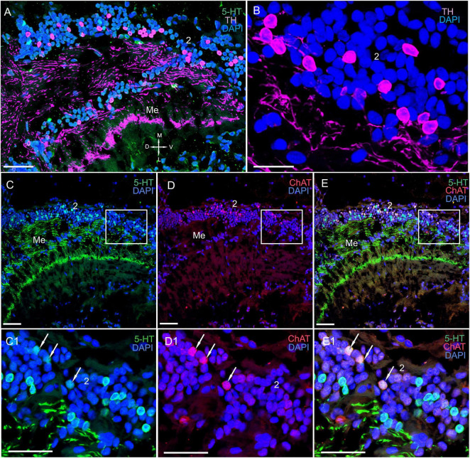
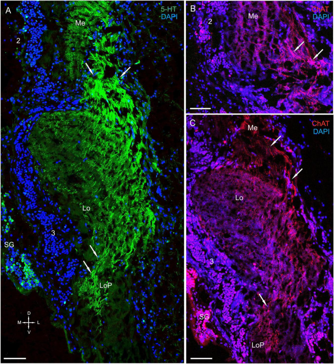
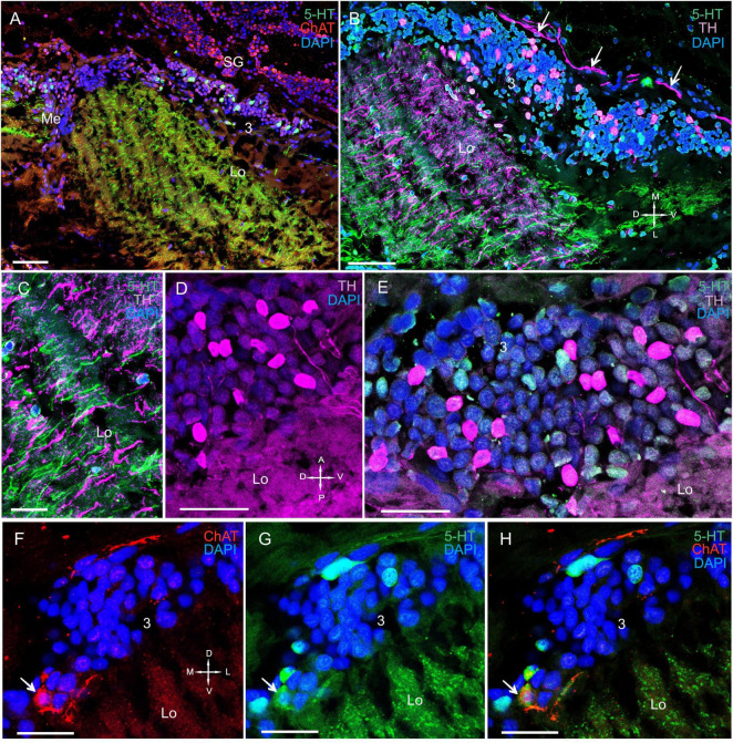
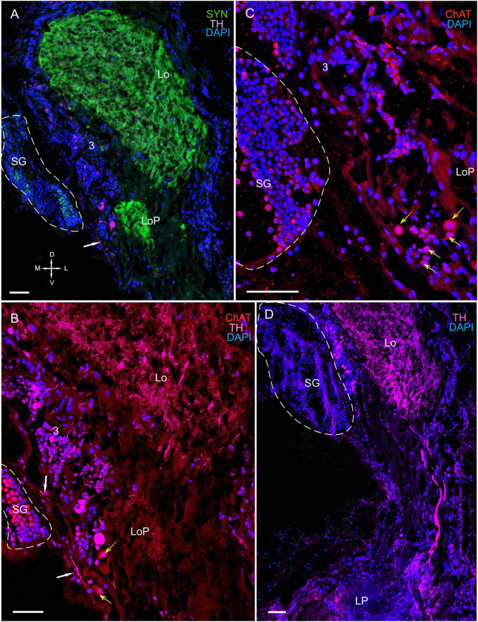
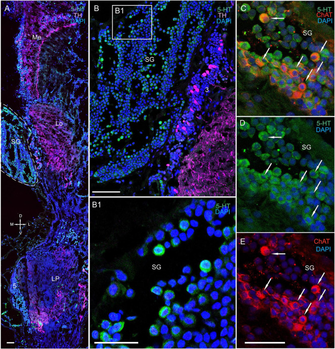
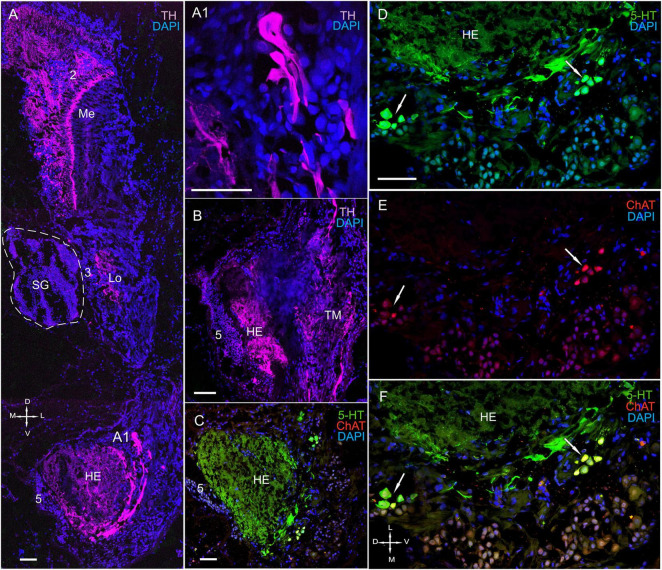
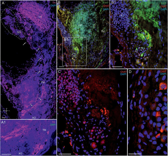
Identifying the neurotransmitters secreted by specific neurons in crustacean eyestalks is crucial to understanding their physiological roles. Here, we combined immunocytochemistry with confocal microscopy and identified the neurotransmitters dopamine (DA), serotonin (5-HT), and acetylcholine (ACh) in the optic neuropils and X-organ sinus gland (XO-SG) complex of the eyestalks of Paralithodes camtschaticus (red king crab). The distribution of Ach neurons was studied by choline acetyltransferase (ChAT) immunohistochemistry and compared with that of DA neurons examined in the same or adjacent sections by tyrosine hydroxylase (TH) immunohistochemistry. We detected 5-HT, TH, and ChAT in columnar, amacrine, and tangential neurons in the optic neuropils and established the presence of immunoreactive fibers and neurons in the terminal medulla in the XO region of the lateral protocerebrum. Additionally, we detected ChAT and 5-HT in the endogenous cells of the SG of P. camtschaticus for the first time. Furthermore, localization of 5-HT- and ChAT-positive cells in the SG indicated that these neurotransmitters locally modulate the secretion of neurohormones that are synthesized in the XO. These findings establish the presence of several neurotransmitters in the XO-SG complex of P. camtschaticus.

Keywords: acetylcholine; crustacea; dopamine; king crab; serotonin; sinus gland; tyrosine hydroxylase.


Conflict of interest statement

The authors declare that the research was conducted in the absence of any commercial or financial relationships that could be construed as a potential conflict of interest.

Figures

FIGURE 1
FIGURE 1
Eyestalks of the red king crab Paralithodes camtschaticus. (A) Photographs demonstrating the position of the eyestalks in P. camtschaticus. (B) Diagram of the optic neuropils, SG, and LP (dorsal view). (C) SYN detected in the optic neuropils and LP of the dorsal to ventral sections. (D) SYN and TH detected in optic neuropils, LP, and OT. Green, SYN; magenta, TH. Dashed lines in panels (C,D) indicate cells corresponding to the SG. Scale bars = (A) 1 cm and (C) 100 μm. La, lamina; Me, medulla; Lo, lobula; LoP, lobula plate; 1, 2, 3, 5, cell clusters; OT, optic tract; LP, lateral protocerebrum; SYN, synapsin; D, dorsal; V, ventral; L, lateral; M, medial; A, anterior; P, posterior.
FIGURE 2
FIGURE 2
Immunolabeled optic neuropils, SG, and lateral protocerebrum. Regions were labeled with DAPI (blue) and anti-SYN (green) or anti-5-HT, anti-ChAT (red), and anti-TH (magenta). (A) Dorsal view of SYN-positive immunostaining in the Me, Lo, and LoP. (B) Ventral view of immunolabeled SYN- and ChAT-positive immunostaining in the Me, Lo, and LoP. (C) Tissue section displaying high TH immunostaining in the LP adjacent to the PT. Dashed line in panel (B) indicates cells of the SG. Scale bars = 100 μm. Me, medulla; Lo, lobula; LoP, lobula plate; LP, lateral protocerebrum; OT, optic tract; PT, protocerebral tract; SYN, synapsin; D, dorsal; V, ventral; L, lateral; M, medial.
FIGURE 3
FIGURE 3
Immunolocalization of 5-HT and ChAT in the lamina of Paralithodes camtschaticus. (A) Horizontal section through an eyestalk showing positive immunostaining for ChAT and 5-HT in the optic neuropils, SG, and LP. (B–D) Detection of ChAT and 5-HT in cell cluster 1 above and below the plexiform layer of the La. (B1–D1) Double immunolabeling of ChAT and 5-HT in cell cluster 1 of the La. Arrows indicate the co-localization of 5-HT with ChAT in neurons. (E) 5-HT-positive immunostaining in varicose processes of the La. Green, 5-HT; red, ChAT; blue, DAPI. Dashed line in panels (A) indicates cells of the SG. Scale bars = (A–D) 100 μm and (B1–E) 50 μm. La, lamina; Me, medulla; Lo, lobula; LP, lateral protocerebrum; 1, 2, 3 cell clusters; OT, optic tract; PT, the protocerebral tract; D, dorsal; V, ventral; L, lateral; M, medial.
FIGURE 4
FIGURE 4
Immunolocalization of TH and 5-HT in the medulla. (A) TH- and 5-HT-positive bodies of neurons connecting the La to the Me. (A1) Localization of TH in neurons at the first Ch and closer to the Me (arrows). (B) Immunolocalization of TH and 5-HT in neurons and nerve fibers of the Me. (C) TH localization in neurons and varicose fibers connecting the La and the Me. Green, 5-HT; magenta, TH; blue, DAPI. Scale bars = (A,B) 100 μm and (A1,C) 50 μm. La, lamina; Me, medulla; 1, 2, cell clusters; Ch, optic chiasma; D, dorsal; V, ventral; A, anterior; P, posterior.
FIGURE 5
FIGURE 5
Immunolocalization of 5-HT, TH, and ChAT in the medulla. (A) TH- and 5-HT-positive neurons and nerve fibers in the Me. (B) Immunohistochemical localization of TH in neurons of cell cluster 2. (C–E) Double immunolabeling of 5-HT and ChAT in neurons and nerve fibers of the Me. (C1–E1) Higher magnification of 5-HT- and ChAT-positive neurons of the Me. Arrows indicate colocalization of 5-HT with ChAT in some of the neurons. Green, 5-HT; red, ChAT; magenta, TH; blue, DAPI. Scale bars = (A,C–E1) 100 μm and (B) 50 μm. Me, medulla; 2, cell clusters; D, dorsal; V, ventral; L, lateral; M, medial.
FIGURE 6
FIGURE 6
Immunolocalization of 5-HT and ChAT in the lobula and the lobula plate. (A) 5-HT-positive nerve fibers in the Lo and LoP. (B) ChAT-positive nerve fibers between in the Me and Lo. (C) ChAT-positive nerve fibers in the Lo and LoP. Arrows indicate immunoreactive nerve fibers. Green, 5-HT; red, ChAT; blue, DAPI. Scale bars = 100 μm. Me, medulla; Lo, lobula; 2, 3 cell clusters; LoP, lobula plate; D, dorsal; V, ventral; L, lateral; M, medial.
FIGURE 7
FIGURE 7
Immunolocalization of 5-HT, TH, and ChAT in the lobula. (A) 5-HT- and ChAT-positive cell bodies and nerve fibers in the Lo. (B) TH- and 5-HT-positive cell bodies and nerve fibers in the Lo with TH-positive fibers running above cell cluster 3. (C) TH-and 5-HT-positive fibers of the Lo. (D) Immunohistochemical localization of TH in the somata of cell cluster 3. (E) Immunohistochemical localization of 5-HT- and TH-positive neurons of cell cluster 3. (F–H) Double immunolabeling of ChAT and 5-HT in cell cluster 3 of the distal part of the Lo. Arrows indicate co-localization of 5-HT with ChAT in neurons. Green, 5-HT; red, ChAT; magenta, TH; blue, DAPI. Scale bars = (A–C) 100 μm and (D–H) 50 μm.
FIGURE 8
FIGURE 8
Immunolocalization of TH, ChAT, and SYN in the lobula, lobula plate, and SG. (A) Photographs demonstrating the position of TH-positive cell bodies near the Lo and LoP. (B) TH- and ChAT-positive immunostaining in the SG (yellow arrows: ChAT-positive somata). TH-positive fibers running between neurons of cluster 3 and the SG (white arrows). (C) ChAT-positive neurons localized near the front edge of the Lo and near the LoP. (D) TH-positive nerve fibers connect the Lo and LP. Green, SYN; magenta, TH; red, ChAT; blue, DAPI. Dashed lines indicate cells of the SG. Scale bars = 100 μm. Lo, lobula; LoP, lobula plate; LP, lateral protocerebrum; SYN, synapsin; D, dorsal; V, ventral; L, lateral; M, medial; A, anterior; P, posterior.
FIGURE 9
FIGURE 9
Immunolocalization of 5-HT, TH, and ChAT in the lobula and SG. (A) Tissue section through an eyestalk of Paralithodes camtschaticus displaying the location of the SG near the Lo. (B) Somata of TH-positive neurons located in cluster 3 near the SG. (B1) Inset (B1) shows 5-HT-positive immunostaining at higher magnification in the endogenous cells of the SG. (C–E) Double immunolabeling of 5-HT and ChAT in cells of the SG and showing co-localization of 5-HT with ChAT in few of the cells (arrows). Green, 5-HT; red, ChAT; magenta, TH; blue, DAPI. Dashed line indicates cells corresponding to the SG. Scale bars = (A,B) 100 μm and (B1–E) 50 μm.
FIGURE 10
FIGURE 10
Immunolocalization of 5-HT, TH, and ChAT in the optic neuropils, SG, and lateral protocerebrum. (A) Tissue section showing high levels of TH-positive immunostaining in the Me, Lo, and LP. (A1) TH-positive cells with large varicose processes in the LP. (B) High levels of TH in processes of both the HE and the TM. (C) ChAT- and 5-HT-positive immunostaining in the HE and neurons in different regions of the TM. (D–F) Double immunolabeling of 5-HT and ChAT in neurons of the TM. Green, 5-HT; red, ChAT; magenta, TH; blue, DAPI. Scale bars = 100 μm. Me, medulla; Lo, lobula; HE, hemiellipsoid body; TM, terminal medulla; PT, protocerebral tract; 2, 3, 5 (ds), cell cluster; D, dorsal; V, ventral; L, lateral; M, medial; A, anterior; P, posterior.
FIGURE 11
FIGURE 11
Immunolocalization of 5-HT, TH, and ChAT in the lateral protocerebrum of Paralithodes camtschaticus. (A,A1) Tissue section through the eyestalk of P. camtschaticus showing TH-positive immunostaining in the Lo and LP. (A1) Regions of the XO showing strong TH immunostaining. (B) TH-positive processes in the LP near the cells of cluster 5 and regions of the TM. (B1–D) 5-HT- and ChAT-positive neurons and processes near cell cluster 5. TH-positive immunostaining in optic neuropils and the LP. Scale bars = 100 μm. Lo, lobula; 5, cell cluster; LP, lateral protocerebrum; TM, terminal medulla; HE, the hemiellipsoid body; D, dorsal; V, ventral; L, lateral; M, medial; A, anterior; P, posterior.

Similar articles

Cited by

References

    1. Allayie S. A., Ravichandran S., Bhat B. A. (2011). Hormonal regulatory role of eyestalk factors on growth of heart in mud crab. Scylla serrata. Saudi J. Biol. Sci. 18 283–286. 10.1016/j.sjbs.2011.02.003 - DOI - PMC - PubMed
    1. Alvarez Alvarado R., Porras, Villalobos M. G., Calderón, Rosete G., Rodríguez-Sosa L., et al. (2005). Dopaminergic modulation of neurosecretory cells in the crayfish. Cell Mol. Neurobiol. 25 345–370. 10.1007/s10571-005-3064-9 - DOI - PMC - PubMed
    1. Andrew R. D., Orchard I., Saleuddin A. S. M. (1978). Structural reevaluation of the neurosecretory system in the crayfish eyestalk. Cell Tissue Res. 190 235–246. 10.1007/BF00218172 - DOI - PubMed
    1. Andrew R. D., Saleuddin A. S. M. (1978). Structure and innervation of a crustacean neurosecretory cell. Can. J. Zool. 56 423–430. 10.1139/z78-060 - DOI
    1. Aréchiga H., Rodriguez-Sosa L. (2002). “Distributed circadian rhythmicity in the crustacean nervous system,” in The Crustacean Nervous System, ed. Wiese K. (Berlin: Springer; ), 113–122. 10.1007/978-3-662-04843-6_8 - DOI

LinkOut - more resources