Review
. 2022 Apr 13;16(2):021502.
doi: 10.1063/5.0084415. eCollection 2022 Mar.

Recent advances in the understanding of alveolar flow

Affiliations
Review

Recent advances in the understanding of alveolar flow

Jun Dong et al. Biomicrofluidics. .

Abstract

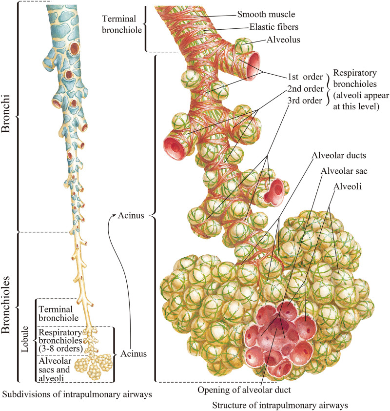
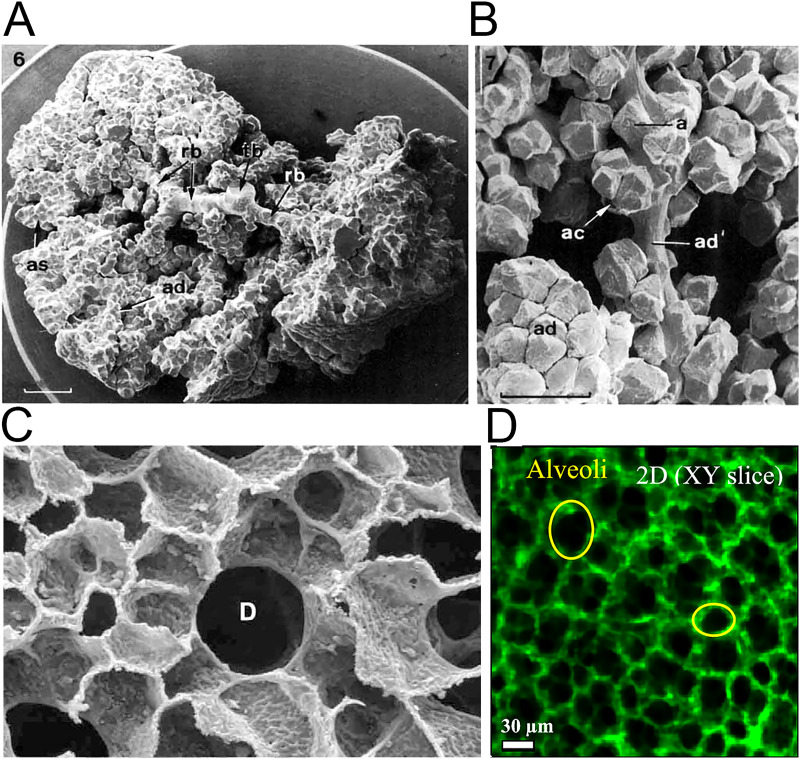
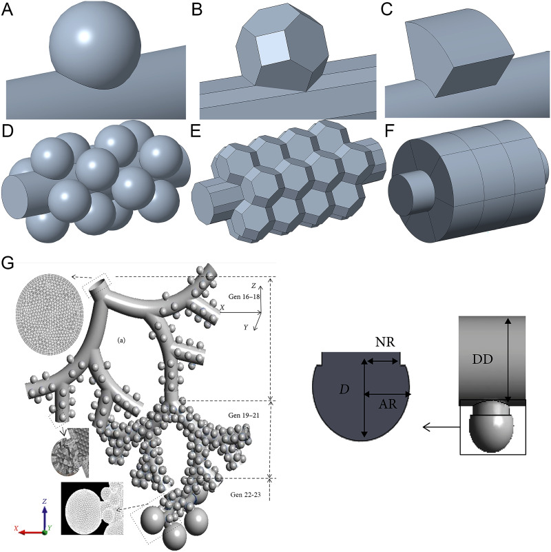
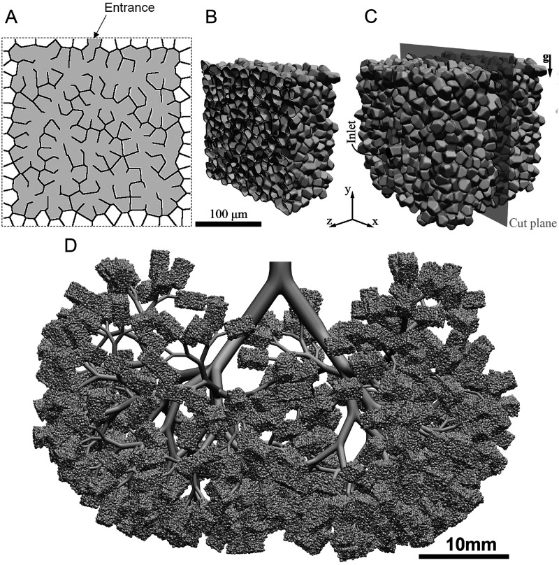
Understanding the dynamics of airflow in alveoli and its effect on the behavior of particle transport and deposition is important for understanding lung functions and the cause of many lung diseases. The studies on these areas have drawn substantial attention over the last few decades. This Review discusses the recent progress in the investigation of behavior of airflow in alveoli. The information obtained from studies on the structure of the lung airway tree and alveolar topology is provided first. The current research progress on the modeling of alveoli is then reviewed. The alveolar cell parameters at different generation of branches, issues to model real alveolar flow, and the current numerical and experimental approaches are discussed. The findings on flow behavior, in particular, flow patterns and the mechanism of chaotic flow generation in the alveoli are reviewed next. The different flow patterns under different geometrical and flow conditions are discussed. Finally, developments on microfluidic devices such as lung-on-a-chip devices are reviewed. The issues of current devices are discussed.


Figures

FIG. 1.
FIG. 1.
Model of a human airway system. From Netter, Atlas of Human Anatomy, 6th Edition. Copyright 2014 Elsevier Inc. Adapted with permission from Elsevier Inc.
FIG. 2.
FIG. 2.
Anatomy of the pulmonary acini and alveoli. (a) Scanning electron micrograph (SEM) of acinus from lung cast by perfusion with formaldehyde. Part of the alveolar ducts (ad) and alveolar sacs (as) were removed to expose the transitional bronchiole (tb) and the respiratory bronchioles (rb); scale bar 1 mm. (b) SEM of portions of sub-acinus. One alveolar duct (ad') is stretched to show the arrangement of single (a) or clustered (ac) alveoli. Dense alveolar accumulation in unstretched alveolar ducts (ad). (c) SEM of the cross section of alveolar duct D surrounded by densely packed alveoli in human lung. (d) Observation of alveoli on a slice of reconstructed three-dimensional rat lung by confocal microscopy technique. Images (a) and (b) are reproduced with permission from Haefeli-Bleuer and Weibel, Anat. Rec. 220(4), 401–414 (1988). Copyright 1988 John Wiley & Sons, Inc. Image (c) is reproduced with permission from Weibel et al., Respir. Physiol. Neurobiol. 148(1–2), 3–21 (2005). Copyright 2005 Elsevier. Image (d) is reproduced with permission from Yang et al., ACS Nano 13(2), 1029–1041 (2019). Copyright 2019 American Chemical Society.
FIG. 3.
FIG. 3.
(a) and (d) spherical alveolus. (b) and (e) Polyhedral (i.e., truncated octahedron) alveolus. (c) and (f) Toroidal alveolus. Images (a)–(c) are single alveolar models. Images (d)–(f) are multi-alveolar models. (g) Alveoli with long necks in whole-lung airway model. From Kolanjiyil and Kleinstreuer, Comput. Math. Method Med. 2019, 5952941 (2019). Copyright 2019 Author(s), licensed under a Creative Commons Attribution (CC BY) license.
FIG. 4.
FIG. 4.
Heterogeneous acinus structure. (a) Two-dimensional schematic diagram of the acinus structure model. (b) Cross-sectional cut through the acinar model. (c) Whole view of the acinar model. (d) Whole-lung model (simplified deep lung model). Image (a) is reproduced with permission from Koshiyama and Wada, Comput. Biol. Med. 62, 25–32 (2015). Copyright 2015 Elsevier Ltd. Images (b) and (c) are reproduced with permission from Hofemeier et al., Eur. J. Pharm. Sci. 113, 53–63 (2018). Copyright 2017 Elsevier B.V. Image (d) is reproduced with permission from Koullapis et al., J. Aerosol. Sci. 144, 105541 (2020). Copyright 2020 Elsevier Ltd.
FIG. 5.
FIG. 5.
Schematic illustrations of control systems for expansion and contraction of alveoli. (a) Alveolar model with alveolar sac. (b) Alveolar model without alveolar sac.
FIG. 6.
FIG. 6.
Models of scaled-up alveoli and alveolar chips. (a) Experimental PIV setup of scaled-up model. (b) Cross section of scaled-up model (magnified 22 times). (c) CAD drawing of multi-generation alveoli chip. (d) Multi-generation alveoli chip. (e) CAD drawing of alveolar chip of single alveolus. (f) Alveolar chip of single alveolus. Images (a) and (b) are reproduced with permission from Berg et al., J. Biomech. 43(6), 1039–1047 (2010). Copyright 2009 Elsevier. Images (c) and (d) are reproduced with permission from Fishler et al., J. Biomech. 46(16), 2817–2823 (2013). Copyright 2013 Elsevier. Images (e) and (f) are reproduced with permission from Lv et al., Lab Chip 20(13), 2394–2402 (2020). Copyright 2020 The Royal Society of Chemistry.
FIG. 7.
FIG. 7.
Images (a)–(h) Flow patterns and velocity fields on the midplane of alveolar cavities at the 16th to 23rd generations. Reproduced with permission from Lv et al., Lab Chip 20(13), 2394–2402 (2020). Copyright 2020 The Royal Society of Chemistry. (i) Recirculation flow pattern with closed streamlines in vortex region (red lines). Reproduced with permission from Henry et al., J. Biomech. Eng. 134(12), 121001 (2012). Copyright 2012 ASME. (j) Schematic diagram of alveoli. θ is the opening half angle, H is the height of alveolar cavity, Qa is alveolar flow rate, Qd is ductal flow rate, Dd is the ductal diameter, and Da is the alveolar diameter (k) Streamlines in an alveolus at birth. Reproduced with permission from Semmler-Behnke et al., Proc. Natl. Acad. Sci. U.S.A. 109(13), 5092–5097 (2012). Copyright 2012 National Academy of Science.
FIG. 8.
FIG. 8.
Chaotic mixing in alveoli. (a) Instantaneous flow patterns with closed streamlines. Reproduced with permission from Tsuda et al., J. Appl. Physiol. 79(3), 1055–1063 (1995). Copyright 1995 American Physiological Society. (b) Instantaneous flow patterns with unclosed streamlines. Reproduced with permission from Lv et al., Lab Chip 20(13), 2394–2402 (2020). Copyright 2020 The Royal Society of Chemistry. (c) Stretched and folded mixing pattern. Reproduced with permission from Butler and Tsuda, J. Appl. Physiol. 83(3), 800–809 (1997). Copyright 1997 American Physiological Society.
FIG. 9.
FIG. 9.
Two- and three-dimensional lung-on-a-chip. (a) The microfabricated lung-on-a-chip device with a two-dimensional alveolar–capillary barrier. Reproduced with permission from Huh et al., Science 328(5986), 1662–1668 (2010). Copyright 2010 American Association for the Advancement of Science. (b) SEM image of spherical microwells (side views; scale bars: 100 μm). Baptista et al., Biomaterials 266, 120436 (2021). Copyright 2021 Author(s), licensed under a Creative Commons Attribution (CC BY) license. (c) Cross-sectional view of three-dimensional alveolar–capillary interface with alveolar epithelial–endothelial/capillary barrier. Baptista et al., Biomaterials 266, 120436 (2021). Copyright 2021, licensed under a Creative Commons Attribution (CC BY) license.

Similar articles

Cited by

References

    1. Chuang H.-C., in Oxidative Stress in Lung Diseases, edited by Chakraborti S., Chakraborti T., Das S. K., and Chattopadhyay D. (Springer, Singapore, 2019), Vol. 1, pp. 293–307.
    1. Tsuda A., Henry F. S., and Butler J. P., Compr. Physiol. 3(4), 1437–1471 (2013). 10.1002/cphy.c100085 - DOI - PMC - PubMed
    1. Hogan B. and Tata P. R., “Cellular organization and biology of the respiratory system,” Nat. Cell Biol. (published online) (2019). 10.1038/s41556-019-0357-7 - DOI - PubMed
    1. Kim K. H., Kabir E., and Kabir S., Environ. Int. 74, 136–143 (2015). 10.1016/j.envint.2014.10.005 - DOI - PubMed
    1. Chen R., Hu B., Liu Y., Xu J. X., Yang G. S., Xu D. D., and Chen C. Y., Biochim. Biophys. Acta Gen. Subj. 1860(12), 2844–2855 (2016). 10.1016/j.bbagen.2016.03.019 - DOI - PubMed

LinkOut - more resources