

Neurobiology of Loneliness, Isolation, and Loss: Integrating Human and Animal Perspectives
- PMID: 35464141
- PMCID: PMC9029604
- DOI: 10.3389/fnbeh.2022.846315
Neurobiology of Loneliness, Isolation, and Loss: Integrating Human and Animal Perspectives
Abstract
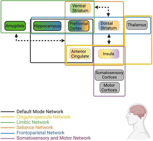
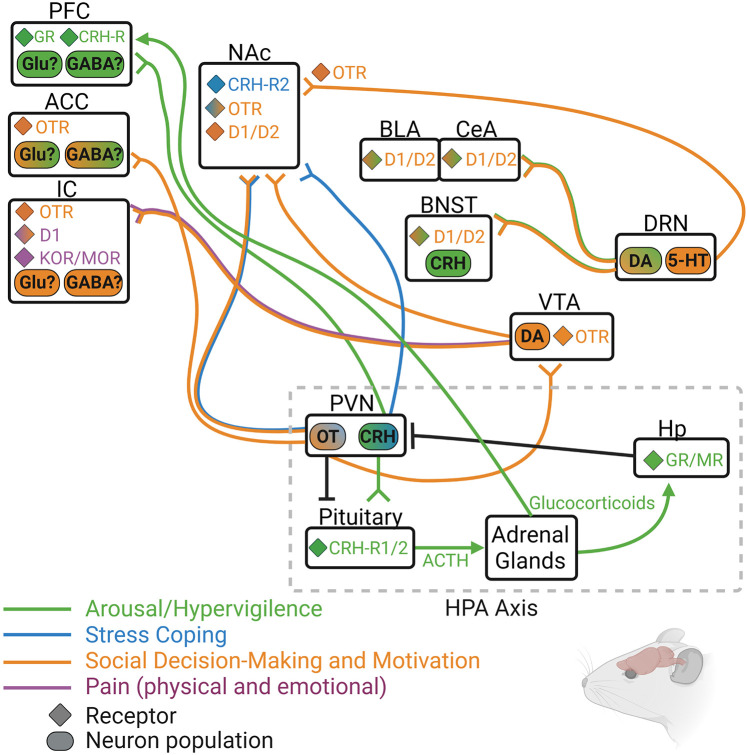
In social species such as humans, non-human primates, and even many rodent species, social interaction and the maintenance of social bonds are necessary for mental and physical health and wellbeing. In humans, perceived isolation, or loneliness, is not only characterized by physical isolation from peers or loved ones, but also involves negative perceptions about social interactions and connectedness that reinforce the feelings of isolation and anxiety. As a complex behavioral state, it is no surprise that loneliness and isolation are associated with dysfunction within the ventral striatum and the limbic system - brain regions that regulate motivation and stress responsiveness, respectively. Accompanying these neural changes are physiological symptoms such as increased plasma and urinary cortisol levels and an increase in stress responsivity. Although studies using animal models are not perfectly analogous to the uniquely human state of loneliness, studies on the effects of social isolation in animals have observed similar physiological symptoms such as increased corticosterone, the rodent analog to human cortisol, and also display altered motivation, increased stress responsiveness, and dysregulation of the mesocortical dopamine and limbic systems. This review will discuss behavioral and neuropsychological components of loneliness in humans, social isolation in rodent models, and the neurochemical regulators of these behavioral phenotypes with a neuroanatomical focus on the corticostriatal and limbic systems. We will also discuss social loss as a unique form of social isolation, and the consequences of bond disruption on stress-related behavior and neurophysiology.
Keywords: CRH; dopamine; isolation; loneliness; loss; opioids; oxytocin.
Copyright © 2022 Vitale and Smith.
Conflict of interest statement
The authors declare that the research was conducted in the absence of any commercial or financial relationships that could be construed as a potential conflict of interest.
Figures

Similar articles
-
Why may allopregnanolone help alleviate loneliness?Med Hypotheses. 2015 Dec;85(6):947-52. doi: 10.1016/j.mehy.2015.09.004. Epub 2015 Sep 5. Med Hypotheses. 2015. PMID: 26365247 Free PMC article.
-
Effects of mental health stigma on loneliness, social isolation, and relationships in young people with depression symptoms.BMC Psychiatry. 2023 Jul 21;23(1):527. doi: 10.1186/s12888-023-04991-7. BMC Psychiatry. 2023. PMID: 37479975 Free PMC article.
-
Affective Neuroscience of Loneliness: Potential Mechanisms underlying the Association between Perceived Social Isolation, Health, and Well-Being.J Psychiatr Brain Sci. 2022;7(6):e220011. doi: 10.20900/jpbs.20220011. Epub 2022 Dec 26. J Psychiatr Brain Sci. 2022. PMID: 36778655 Free PMC article.
-
Neural mechanisms of social homeostasis.Ann N Y Acad Sci. 2019 Dec;1457(1):5-25. doi: 10.1111/nyas.14016. Epub 2019 Mar 15. Ann N Y Acad Sci. 2019. PMID: 30875095 Free PMC article. Review.
-
[Neurobiology of Loneliness, Social Isolation, and Affiliative Social Behavior].Brain Nerve. 2023 Mar;75(3):263-268. doi: 10.11477/mf.1416202318. Brain Nerve. 2023. PMID: 36890762 Review. Japanese.
Cited by
-
Can social isolation alleviate symptoms of anxiety and depression disorders?Front Psychiatry. 2025 Apr 16;16:1561916. doi: 10.3389/fpsyt.2025.1561916. eCollection 2025. Front Psychiatry. 2025. PMID: 40309500 Free PMC article. Review.
-
Loneliness and social conformity: A predictive processing perspective.Ann N Y Acad Sci. 2025 May;1547(1):5-17. doi: 10.1111/nyas.15324. Epub 2025 Apr 2. Ann N Y Acad Sci. 2025. PMID: 40173107 Free PMC article. Review.
-
Association between Migraine and Workplace Social Support in the Social Context of China: Using a Validated Chinese Version of the DCSQ.Healthcare (Basel). 2023 Jan 5;11(2):171. doi: 10.3390/healthcare11020171. Healthcare (Basel). 2023. PMID: 36673539 Free PMC article.
-
The Impact of Loneliness and Social Isolation on Cognitive Aging: A Narrative Review.J Alzheimers Dis Rep. 2023 Jun 29;7(1):699-714. doi: 10.3233/ADR-230011. eCollection 2023. J Alzheimers Dis Rep. 2023. PMID: 37483321 Free PMC article. Review.
-
Breaking the vicious cycle: The interplay between loneliness, metabolic illness, and mental health.Front Psychiatry. 2023 Mar 8;14:1134865. doi: 10.3389/fpsyt.2023.1134865. eCollection 2023. Front Psychiatry. 2023. PMID: 36970267 Free PMC article.
References
-
- Anderson C. M., Martin M. M. (1995). The effects of communication motives, interaction involvement, and loneliness on satisfaction: a model of small groups. Small Group Res. 26 118–137. 10.1177/1046496495261007 - DOI
Publication types
Grants and funding
LinkOut - more resources
Full Text Sources
Research Materials