A Neuroanatomy of Positive Affect Display - Subcortical Fiber Pathways Relevant for Initiation and Modulation of Smiling and Laughing
- PMID: 35464145
- PMCID: PMC9022623
- DOI: 10.3389/fnbeh.2022.817554
A Neuroanatomy of Positive Affect Display - Subcortical Fiber Pathways Relevant for Initiation and Modulation of Smiling and Laughing
Abstract
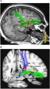
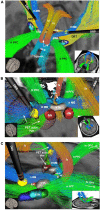
Background: We here report two cases of stimulation induced pathological laughter (PL) under thalamic deep brain stimulation (DBS) for essential tremor and interpret the effects based on a modified neuroanatomy of positive affect display (PAD).
Objective/hypothesis: The hitherto existing neuroanatomy of PAD can be augmented with recently described parts of the motor medial forebrain bundle (motorMFB). We speculate that a co-stimulation of parts of this fiber structure might lead to a non-volitional modulation of PAD resulting in PL.
Methods: We describe the clinical and individual imaging workup and combine the interpretation with normative diffusion tensor imaging (DTI)-tractography descriptions of motor connections of the ventral tegmental area (VTA) (n = 200 subjects, HCP cohort), [[18F] fluorodeoxyglucose (18FDG)] positron emission tomography (PET), and volume of activated tissue simulations. We integrate these results with literature concerning PAD and the neuroanatomy of smiling and laughing.
Results: DBS electrodes bilaterally co-localized with the MB-pathway ("limiter pathway"). The FDG PET activation pattern allowed to explain pathological PAD. A conceptual revised neuroanatomy of PAD is described.
Conclusion: Eliciting pathological PAD through chronic thalamic DBS is a new finding and has previously not been reported. PAD is evolution driven, hard wired to the brain and realized over previously described branches of the motorMFB. A major relay region is the VTA/mammillary body complex. PAD physiologically undergoes conscious modulation mainly via the MB branch of the motorMFB (limiter). This limiter in our cases is bilaterally disturbed through DBS. The here described anatomy adds to a previously described framework of neuroanatomy of laughter and humor.
Keywords: deep brain stimulation; essential tremor; laughter; motorMFB; pathological laughter; pseudobulbar affect.
Copyright © 2022 Coenen, Sajonz, Hurwitz, Böck, Hosp, Reinacher, Urbach, Blazhenets, Meyer and Reisert.
Conflict of interest statement
The authors declare that the research was conducted in the absence of any commercial or financial relationships that could be construed as a potential conflict of interest.
Figures


Similar articles
-
Tractography-assisted deep brain stimulation of the superolateral branch of the medial forebrain bundle (slMFB DBS) in major depression.Neuroimage Clin. 2018 Aug 14;20:580-593. doi: 10.1016/j.nicl.2018.08.020. eCollection 2018. Neuroimage Clin. 2018. PMID: 30186762 Free PMC article. Clinical Trial.
-
Ventral tegmental area connections to motor and sensory cortical fields in humans.Brain Struct Funct. 2019 Nov;224(8):2839-2855. doi: 10.1007/s00429-019-01939-0. Epub 2019 Aug 22. Brain Struct Funct. 2019. PMID: 31440906 Free PMC article.
-
Mapping Motor Pathways in Parkinson's Disease Patients with Subthalamic Deep Brain Stimulator: A Diffusion MRI Tractography Study.Neurol Ther. 2022 Jun;11(2):659-677. doi: 10.1007/s40120-022-00331-1. Epub 2022 Feb 14. Neurol Ther. 2022. PMID: 35165822 Free PMC article.
-
Deep brain stimulation of the brainstem.Brain. 2021 Apr 12;144(3):712-723. doi: 10.1093/brain/awaa374. Brain. 2021. PMID: 33313788 Free PMC article.
-
Diffusion Tractography in Deep Brain Stimulation Surgery: A Review.Front Neuroanat. 2016 May 2;10:45. doi: 10.3389/fnana.2016.00045. eCollection 2016. Front Neuroanat. 2016. PMID: 27199677 Free PMC article. Review.
Cited by
-
Deconstructing a common pathway concept for Deep Brain Stimulation in the case of Obsessive-Compulsive Disorder.Mol Psychiatry. 2025 Sep;30(9):4274-4285. doi: 10.1038/s41380-025-03008-x. Epub 2025 Apr 6. Mol Psychiatry. 2025. PMID: 40189699 Free PMC article.
-
"The Heart Asks Pleasure First"-Conceptualizing Psychiatric Diseases as MAINTENANCE Network Dysfunctions through Insights from slMFB DBS in Depression and Obsessive-Compulsive Disorder.Brain Sci. 2022 Mar 25;12(4):438. doi: 10.3390/brainsci12040438. Brain Sci. 2022. PMID: 35447971 Free PMC article.
-
The anterior perforated substance (APS) revisited: Commented anatomical and imagenological views.Brain Behav. 2023 Dec;13(12):e3029. doi: 10.1002/brb3.3029. Epub 2023 Nov 27. Brain Behav. 2023. PMID: 38010896 Free PMC article.
References
-
- Coenen V. A., Döbrössy M. D., Teo S. J., Wessolleck J., Sajonz B. E. A., Reinacher P. C., et al. (2021). Diverging prefrontal cortex fiber connection routes to the subthalamic nucleus and the mesencephalic ventral tegmentum investigated with long range (normative) and short range (ex-vivo high resolution) 7T DTI. Brain Struct. Funct. 2021 1–25. 10.1007/s00429-021-02373-x - DOI - PMC - PubMed
-
- Coenen V. A., Honey C. R., Hurwitz T., Rahman A. A., McMaster J., Bürgel U., et al. (2009). Medial forebrain bundle stimulation as a pathophysiological mechanism for hypomania in subthalamic nucleus deep brain stimulation for parkinson’s disease. Neurosurgery 64 1106–1114. 10.1227/01.neu.0000345631.54446.06 - DOI - PubMed
-
- Coenen V. A., Panksepp J., Hurwitz T. A., Urbach H., Mädler B. (2012). Human Medial Forebrain Bundle (MFB) and Anterior Thalamic Radiation (ATR): imaging of two major subcortical pathways and the dynamic balance of opposite affects in understanding depression. J. Neuropsych. Clin. Neurosci. 24 223–236. 10.1176/appi.neuropsych.11080180 - DOI - PubMed
LinkOut - more resources
Full Text Sources
Miscellaneous

