

Age-related functional decline of human B cells
- PMID: 35464165
- PMCID: PMC8975901
- DOI: 10.1007/s10616-021-00513-z
Age-related functional decline of human B cells
Abstract
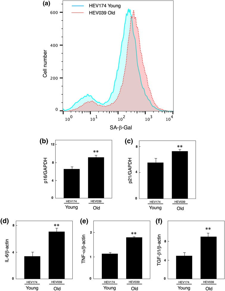
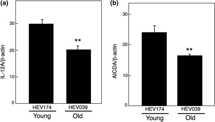
This study aimed to investigate the changes in B cell functional decline and antigen sensitization with aging using two Epstein Barr virus (EBV)-immortalized human B cell lines, one from a 22-year-old man (EBV-B young) and the other from a 65-year-old man (EBV-B old). The activity of senescence-associated β-galactosidase, a marker of cellular senescence, was enhanced in the EBV-B old cells compared with EBV-B young cells. Moreover, the levels of p16, p21, IL-6, TNF-α, and TGF-β1, which are senescence-associated secretary phenotypes, were also increased in EBV-B old cells. In vitro immunization of EBV-B cells with β-lactoglobulin further showed that EBV-B old cells had a reduced cell population of naïve B cells than that of EBV-B young cells. Furthermore, HLA-DR expression, which is important for antigen presentation, was decreased in the EBV-B old cells. Comparative microarray analysis between EBV-B young and old cells also showed decreased expression of antibody genes, such as those of the heavy chain and light chain (κ chain). These results suggest that cellular senescence and decreased gene expression are responsible, at least in part, for the decline in B cell function and antigen sensitization capacity with aging, which ultimately impairs the function of the acquired immune system.
Keywords: Antigen sensitization; Epstein Barr virus-immortalized B cell; Immune senescence; Senescence-associated secretory phenotype.
© The Author(s), under exclusive licence to Springer Nature B.V. 2021.
Conflict of interest statement
Conflict of interestThe authors have no relevant financial or non-financial interests to disclose.
Figures


Similar articles
-
BZLF1 Attenuates Transmission of Inflammatory Paracrine Senescence in Epstein-Barr Virus-Infected Cells by Downregulating Tumor Necrosis Factor Alpha.J Virol. 2016 Aug 12;90(17):7880-93. doi: 10.1128/JVI.00999-16. Print 2016 Sep 1. J Virol. 2016. PMID: 27334596 Free PMC article.
-
Molecular characterization of major histocompatibility complex class II gene expression and demonstration of antigen-specific T cell response indicate a new phenotype in class II-deficient patients.J Exp Med. 1995 Apr 1;181(4):1411-23. doi: 10.1084/jem.181.4.1411. J Exp Med. 1995. PMID: 7699327 Free PMC article.
-
Expression of mucin (MUC-1) from a mini-Epstein-Barr virus in immortalized B-cells to generate tumor antigen specific cytotoxic T cells.J Gene Med. 1999 Mar-Apr;1(2):84-92. doi: 10.1002/(SICI)1521-2254(199903/04)1:2<84::AID-JGM21>3.0.CO;2-Q. J Gene Med. 1999. PMID: 10738572
-
Age-related EBV-associated B-cell lymphoproliferative disorders and other EBV + lymphoproliferative diseases: New insights into immune escape and immunodeficiency through staining with anti-PD-L1 antibody clone SP142.Pathol Int. 2020 Aug;70(8):481-492. doi: 10.1111/pin.12946. Epub 2020 May 4. Pathol Int. 2020. PMID: 32367595 Review.
-
Interactions involving cyclosporine A, interleukin-6, and Epstein-Barr virus lead to the promotion of B-cell lymphoproliferative disease.Leuk Lymphoma. 1996 May;21(5-6):379-90. doi: 10.3109/10428199609093435. Leuk Lymphoma. 1996. PMID: 9172802 Review.
Cited by
-
Age-associated changes in innate and adaptive immunity: role of the gut microbiota.Front Immunol. 2024 Sep 16;15:1421062. doi: 10.3389/fimmu.2024.1421062. eCollection 2024. Front Immunol. 2024. PMID: 39351234 Free PMC article. Review.
-
Impact of Particulate Matter Exposure and Aerobic Exercise on Circulating Biomarkers of Oxidative Stress, Antioxidant Status, and Inflammation in Young and Aged Mice.Life (Basel). 2023 Sep 23;13(10):1952. doi: 10.3390/life13101952. Life (Basel). 2023. PMID: 37895334 Free PMC article.
-
Senescent macrophages in cancer: roles in tumor progression and treatment opportunities.Cancer Biol Med. 2025 May 6;22(5):439-59. doi: 10.20892/j.issn.2095-3941.2024.0589. Cancer Biol Med. 2025. PMID: 40329498 Free PMC article. Review.
References
LinkOut - more resources
Full Text Sources
Research Materials
