Review
. 2022 Apr 7:13:818736.
doi: 10.3389/fimmu.2022.818736. eCollection 2022.

Sialylation as an Important Regulator of Antibody Function

Affiliations
Review

Sialylation as an Important Regulator of Antibody Function

Ravi Vattepu et al. Front Immunol. .

Abstract

Antibodies play a critical role in linking the adaptive immune response to the innate immune system. In humans, antibodies are categorized into five classes, IgG, IgM, IgA, IgE, and IgD, based on constant region sequence, structure, and tropism. In serum, IgG is the most abundant antibody, comprising 75% of antibodies in circulation, followed by IgA at 15%, IgM at 10%, and IgD and IgE are the least abundant. All human antibody classes are post-translationally modified by sugars. The resulting glycans take on many divergent structures and can be attached in an N-linked or O-linked manner, and are distinct by antibody class, and by position on each antibody. Many of these glycan structures on antibodies are capped by sialic acid. It is well established that the composition of the N-linked glycans on IgG exert a profound influence on its effector functions. However, recent studies have described the influence of glycans, particularly sialic acid for other antibody classes. Here, we discuss the role of glycosylation, with a focus on terminal sialylation, in the biology and function across all antibody classes. Sialylation has been shown to influence not only IgG, but IgE, IgM, and IgA biology, making it an important and unappreciated regulator of antibody function.

Keywords: Immunoglobulins; antibody; glycosylation; in vitro glycoengineering; in vivo glyco engineering; sialic acid; sialylation.


Conflict of interest statement

The authors declare that the research was conducted in the absence of any commercial or financial relationships that could be construed as a potential conflict of interest.

Figures

Figure 1
Figure 1
Schematic representations of human antibody structures and attached glycans. (A) Human immunoglobulin structures with corresponding glycosylation sites (human numbering) are shown. Each antibody isotype is comprised identical heavy chains (gray) and light chains (white), forming the antigen-binding fragment (Fab) and fragment crystallizable region (Fc). While monomeric structures are shown, IgM typically multimerizes into a pentamers or hexamers, and IgA1 and IgA2 can form dimers. Blue hexagons denote sites occupied by complex-type N-glycans. Green triangles indicate sites occupied by oligomannose N-glycans. Yellow circles mark sites occupied by O-linked glycans. The N383 site on human IgE is unoccupied, and N207 on human IgA2 is present only in specific allotypes. (B) Structures of N- and O-linked glycans found on human antibodies are represented. N-linked glycans are attached to asparagine residues containing the consensus sequence N-X-S/T. N-linked glycans can form complex, high-mannose, or hybrid types. Blue squares; GlcNAc. Yellow circles; galactose. Green circles; mannose. Red triangles; fucose. Yellow squares; GalNAc. Purple diamonds; sialic acid. Dashed lines indicate core structures, and sugar residues outside the core are variable additions.
Figure 2
Figure 2
The impact of N- and O-linked sialylation across antibody classes. Antibody schematics are shown, along with skeleton sialylated (terminating in purple diamonds) or asialylated N-linked glycans. O-glycans and IgA1 are comprised of GalNAc (Yellow squares), galactose (yellow circles), and sialic acid (purple diamonds).
Figure 3
Figure 3
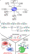
Glycoengineering methods in production cell lines (A), in vitro (B), and in vivo (C). (A) Various genetic manipulations have been performed in production cells to produce differentially fucosylated antibodies. In some cell lines, such as GMD and FX knockouts, fucosylation can be rescued by the addition of substrates such as L-fucose. (B) In vitro methods of glycoengineering and sialylation include step-wise enzymatic building of glycans and chemoenzymatic engineering. (C) In vivo glycoengineering involves administration of enzymes, and have had success in some pre-clinical animal models and on human cells.

References

    1. Imbach P. Treatment of Immune Thrombocytopenia With Intravenous Immunoglobulin and Insights for Other Diseases. A Historical Review. Swiss Med Wkly (2012) 142:w13593. doi: 10.4414/smw.2012.13593 - DOI - PubMed
    1. Bayry J, Negi VS, Kaveri SV. Intravenous Immunoglobulin Therapy in Rheumatic Diseases. Nat Rev Rheumatol (2011) 7(6):349–59. doi: 10.1038/nrrheum.2011.61 - DOI - PubMed
    1. Schwab I, Nimmerjahn F. Intravenous Immunoglobulin Therapy: How Does IgG Modulate the Immune System? Nat Rev Immunol (2013) 13(3):176–89. doi: 10.1038/nri3401 - DOI - PubMed
    1. Mullard A. FDA Approves 100th Monoclonal Antibody Product. Nat Rev Drug Discovery (2021) 20(7):491–5. doi: 10.1038/d41573-021-00079-7 - DOI - PubMed
    1. Harris LJ, Larson SB, Hasel KW, Day J, Greenwood A, McPherson A. The Three-Dimensional Structure of an Intact Monoclonal Antibody for Canine Lymphoma. Nature (1992) 360(6402):369–72. doi: 10.1038/360369a0 - DOI - PubMed