Case Reports
. 2022 Apr 12;17(6):2081-2084.
doi: 10.1016/j.radcr.2022.02.035. eCollection 2022 Jun.

Posterior cerebral artery dissection after excessive caffeine consumption in a teenager

Affiliations
Case Reports

Posterior cerebral artery dissection after excessive caffeine consumption in a teenager

Nikolaos Staikoglou et al. Radiol Case Rep. .

Abstract

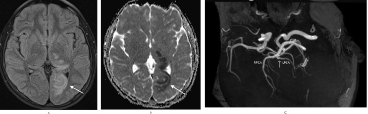
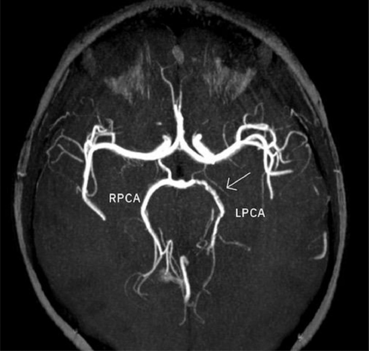
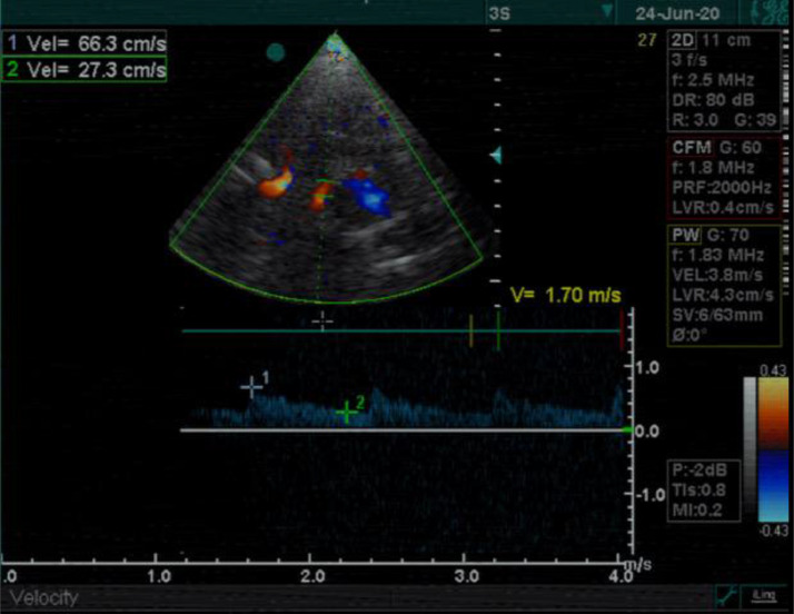
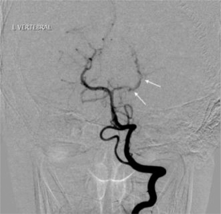
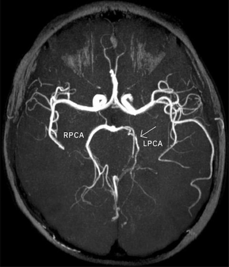
Arterial ischemic stroke is a rare but significant cause of neurological deficits in childhood. Even though there is a variety of risk factors, identifying the etiology can sometimes be a hard diagnostic challenge. Arteriopathies in general, and more specifically, arterial dissection is one of the uncommon pathologies that can cause incidents of pediatric stroke. We report a rare case of a young adolescent with posterior cerebral artery dissection after excessive consumption of caffeine, contained in energy drinks, only hours before the onset of neurological symptoms. A complete neuroimaging evaluation (MRI, intracranial US and digital subtraction angiography) at the admission and during the follow-ups supported the diagnosis of arterial dissection possibly caused by caffeine overconsumption.

Keywords: Caffeine; Dissection; Pediatric stroke; Posterior cerebral artery.


Figures

Fig 1
Fig. 1
MRI on Day 1. (A) There exists a high signal in the cortex of the inner surface of the left occipital lobe and in the left thalamus (arrow). (B) The lesions have a low signal on the ADC map characteristic for ischemia. C) On a 3D-TOF oblique projection, there is a narrowing at the P1-P2 junction while the P4 segment is not apparent.
Fig 2
Fig. 2
Imaging on Day 4 shows the typical “string and pearl” sign of the left P2 and P3 PCA segments on 3D-TOF sequences.
Fig 3
Fig. 3
Intracranial Doppler ultrasonographic examination at the level of the left P1-P2 segment shows an attenuated flow pattern compared to the contralateral side (not shown).
Fig 4
Fig. 4
Digital subtraction angiography on Day 11 demonstrates the typical “string and pearl” sign at the level of the left P1-P2 junction and distally.
Fig 5
Fig. 5
Imaging on day 30 shows residual narrowing of the left PCA.

References

    1. deVeber G.A., Kirton A., Booth F., Yager J., Wirrell E., Wood E., et al. Epidemiology and outcomes of arterial ischemic stroke in children: the canadian pediatric ischemic stroke registry. Pediatr. Neurol. 2017;69:58–70. doi: 10.1016/j.pediatrneurol.2017.01.016. - DOI - PubMed
    1. Bernard T., Manco-Johnson M., Warren L., MacKay M., Ganesan V., DeVeber G., et al. Towards a consensus-based classification of childhood arterial ischemic stroke. Stroke. 2012;43(2):371–377. doi: 10.1161/STROKEAHA.111.624585. - DOI - PMC - PubMed
    1. Lo W., Khaled Z., Ponnappa K., Allen A., Chisolm D., Tang M., et al. The Cost of Pediatric Stroke Care and Rehabilitation. Stroke. 2008;39(1):161–165. doi: 10.1161/STROKEAHA.107.497420. - DOI - PubMed
    1. Mardee G., Anne G., Vicki A., T M.M. Outcome in Childhood Stroke. Stroke. 2016;47(4):1159–1164. doi: 10.1161/STROKEAHA.115.011622. - DOI - PubMed
    1. Mallick A., Ganesan V., Kirkham F., Fallon P., Hedderly T., McShane T., et al. Childhood arterial ischemic stroke incidence, presenting features, and risk factors: a prospective population-based study. Lancet Neurol. 2014;13(1):35–43. doi: 10.1016/S1474-4422(13)70290-4. - DOI - PubMed

Publication types

LinkOut - more resources