Case Reports
. 2022 Apr 12;17(6):2111-2114.
doi: 10.1016/j.radcr.2022.03.072. eCollection 2022 Jun.

Medial tentorial dural arteriovenous fistula: A rare cause of bithalamic oedema

Affiliations
Case Reports

Medial tentorial dural arteriovenous fistula: A rare cause of bithalamic oedema

Cyril Dargazanli et al. Radiol Case Rep. .

Abstract

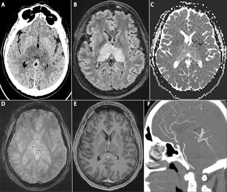
A 39-year-old man was admitted after 1 week of headaches and cognitive changes. CT scan showed bithalamic hypodensities, corresponding to bithalamic vasogenic oedema. Punctuate hemorrhage was present, with foci of thalamic enhancement. CT angiography raised the suspicion of arteriovenous shunt. Digital subtraction angiography confirmed a medial falcotentorial dural arteriovenous fistula. Complete embolization was performed using liquid embolic agent. Although tentorial dural fistulas have already been described as a cause of bithalamic oedema and subacute dementia, they are not generally included in pathologies implied in this radiologic pattern.

Keywords: Bithalamic oedema; Dural arteriovenous fistula; Embolization; Fistula; Neuroradiology.


Figures

Fig 1
Fig. 1
Noninvasive Imaging A: Plain CT scan showing bithalamic symmetrical hypodensities B: Baseline MRI showing bithalamic FLAIR hyperintensities C: Diffusion MRI depicting increased bithalamic apparent diffusion coefficient (ADC) D: Punctuate hemorrhage in right thalamus on T2* E: Gadolinium enhanced sequence showing small foci of bithalamic patchy enhancement F: Arterialized superior vermian vein (white arrow) draining into vein of Galen (dotted white arrow) and subsequently into internal cerebral vein (black arrow).
Fig 2
Fig. 2
Digital Subtraction Angiography (DSA) A and B: Lateral (A) and anteroposterior (B) arterial phase showing the fistulous plaque (white arrow), the arterialized superior vermian vein (dotted white arrow) and vein of Galen (black arrow) C and D: Small contribution of the medial tentorial branch from the left middle meningeal artery (white arrow) E: Lateral venous phase of left vertebral artery injection showing filling defect of the anterior part of straight sinus, related to deep venous thrombosis F: Post embolization non-substracted image showing liquid-embolic cast in the arterial feeders, in the fistulous plaque, but away from the vein of Galen.

References

    1. Holekamp TF, Mollman ME, Murphy RKJ, Kolar GR, Kramer NM, Derdeyn CP, et al. Dural arteriovenous fistula-induced thalamic dementia: report of 4 cases. J Neurosurg. 2016;124(6):1752–1765. doi: 10.3171/2015.5.JNS15473. - DOI - PubMed
    1. Cox M, Rodriguez P, Mohan S, Sedora-Roman NI, Pukenas B, Choudhri O, et al. Tentorial dural arteriovenous fistulas as a cause of thalamic edema: 2 cases of an important differential diagnosis to consider. The Neurohospitalist. 2021;11(1):33–39. doi: 10.1177/1941874420944333. - DOI - PMC - PubMed
    1. Hegde AN, Mohan S, Lath N, Lim CCT. Differential diagnosis for bilateral abnormalities of the basal ganglia and thalamus. RadioGraphics. 2011;31(1):5–30. doi: 10.1148/rg.311105041. - DOI - PubMed
    1. Lawton MT, Sanchez-Mejia RO, Pham D, Tan J, Halbach VV. Tentorial dural arteriovenous fistulae: operative strategies and microsurgical results for six types. Oper Neurosurg. 2008;62(suppl_1) doi: 10.1227/01.neu.0000317381.68561.b0. ONS110-ONS125. - DOI - PubMed

Publication types

LinkOut - more resources