. 2022 May 19;2(5):1084-1099.
doi: 10.1016/j.checat.2022.03.019. Epub 2022 Apr 19.

Mechanism of reaction of RNA-dependent RNA polymerase from SARS-CoV-2

Affiliations

Mechanism of reaction of RNA-dependent RNA polymerase from SARS-CoV-2

Juan Aranda et al. Chem Catal. .

Abstract

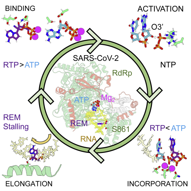
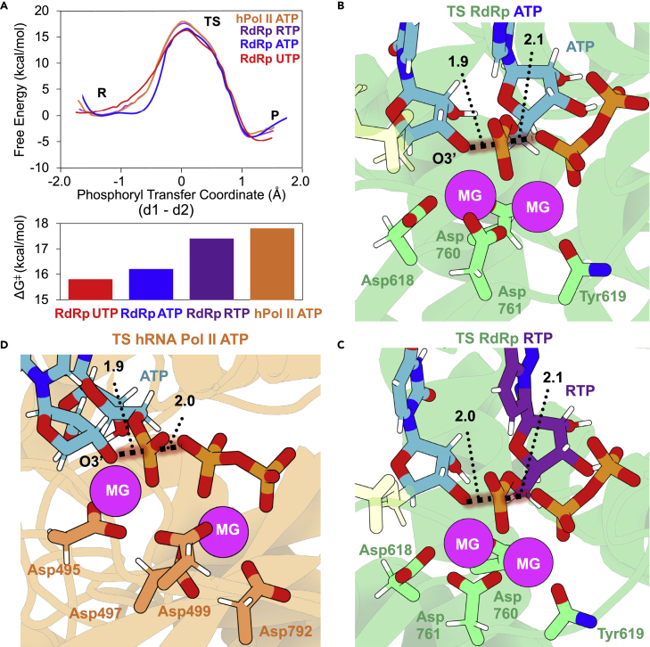
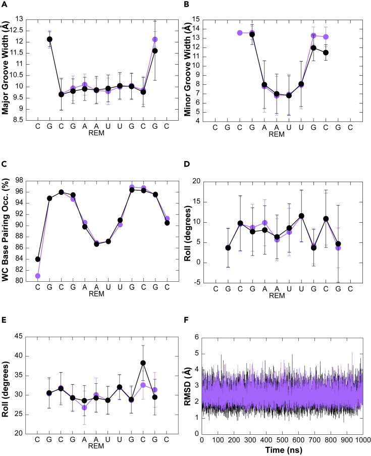
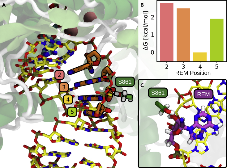
We combine molecular dynamics, statistical mechanics, and hybrid quantum mechanics/molecular mechanics simulations to describe mechanistically the severe acute respiratory syndrome coronavirus 2 (SARS-CoV-2) RNA-dependent RNA polymerase (RdRp). Our study analyzes the binding mode of both natural triphosphate substrates as well as remdesivir triphosphate (the active form of drug), which is bound preferentially over ATP by RdRp while being poorly recognized by human RNA polymerase II (RNA Pol II). A comparison of incorporation rates between natural and antiviral nucleotides shows that remdesivir is incorporated more slowly into the nascent RNA compared with ATP, leading to an RNA duplex that is structurally very similar to an unmodified one, arguing against the hypothesis that remdesivir is a competitive inhibitor of ATP. We characterize the entire mechanism of reaction, finding that viral RdRp is highly processive and displays a higher catalytic rate of incorporation than human RNA Pol II. Overall, our study provides the first detailed explanation of the replication mechanism of RdRp.

Keywords: QM/MM; RNA-dependent RNA polymerase; SARS-CoV-2; antivirals; biocatalysis; free energy calculations; reaction mechanism; remdesivir; viral replication.


Conflict of interest statement

The authors declare no competing interests.

Figures

None
Graphical abstract
Figure 1
Figure 1
Active site of SARS-CoV-2 RdRp makes it an efficient polymerase (A) The replication complex of SARS-CoV-2 formed by the nsp12 RdRp enzyme (in green), the nsp8 and nsp7 cofactors (orange and gray, respectively), and an RNA template and nascent strands (yellow). (B) Scheme depicting the two-metal-ion mechanism used by SARS-CoV-2 RdRp. (C) The active site contains a well-defined coordination sphere of the two Mg2+ ions. (D) NTP substrate recognition in the active site of RdRp is mediated by a pair of arginines, an aspartate, and a serine. (E) The deprotonated 3′ terminal nucleotide is stabilized by one of the catalytic ions in the RdRp’s active site. (F) RdRp active site pocket enables base specificity between the incoming nucleotide and the template. Figures were prepared with 3D Protein Imager. A 3D structure representation can be accessed through https://mmb.irbbarcelona.org/3dRS/s/Y17cub.
Figure 2
Figure 2
Binding preferences in viral and human RNA polymerases (A) Binding free energies of ATP and RTP inside SARS-CoV-2 RdRp and human RNA Pol II (negative values corresponding to a preference for RTP over ATP). (B) Most important interactions between RTP nitrile group and RdRp polymerase. (C) RTP inside human RNA Pol II active site.
Figure 3
Figure 3
Mechanism of activation through O3′ deprotonation inside RdRp (A) Scheme depicting the activation mechanism used by SARS-CoV-2 RdRp. (B) Free-energy profile as a function of the proton transfer coordinate (d3-d4), obtained for the proton transfer reaction between the O3′ of the just-incorporated nucleotide and the γ phosphate group of the newly formed PPi. (C) Transition state (TS) where the H3′ proton belonging to O3′ atom of nucleotide “i” is halfway to being transferred to γ phosphate group of PPi. Reaction coordinate (RC) consisted of the distances between O3′ or Oγ atoms to the H atom to be transferred, displayed as d3 and d4, respectively.
Figure 4
Figure 4
RNA elongation inside RdRp of SARS-CoV-2 and human RNA Pol II (A) Top, free-energy profiles as a function of the phosphoryl transfer coordinate (d1-d2) for the incorporation of a U, an A, or an R to a nascent viral RNA strand. Profile for the incorporation of the inside human RNA Pol II is also shown. The phosphorylation reaction consists of a nucleophilic attack of the O3′ of the terminal nucleotide on the Pα of the triphosphate nucleotide. Bottom, bar plot displaying free energies of activation for the process for different NTPs and enzymes. (B and C) Active site views of the TSs were found when ATP (B) and RTP (C) (purple C atoms) are the substrates for the elongation reaction inside RdRp. The phosphoryl group is half-way to being transferred. One Mg2+ activates the O3′ toward nucleophilic attack and stabilizes the negatively charged TS. The other Mg2+ stabilizes the charged TS, as well as the nascent negatively charged PPi molecule. Distances involved in the reaction are shown as dotted lines with their average values in Å. (D) TS insight of the elongation reaction catalyzed by human RNA pol II.
Figure 5
Figure 5
Remdesivir does not distort RNA structure (A–F) Major (A) and minor (B) groove width, Watson-Crick base pairing (C), roll (D), twist (E), and RMSD (F) of the double helix are not affected when remdesivir is present (purple) with respect to control sequence (black) during MD simulations. The average values across the simulations are shown in black and purple dots for the control and remdesivir-containing sequences, respectively. Average standard deviations are shown as black and purple bars. The RMSD of one of the five replicas is shown in (F) (see also Figure S11).
Figure 6
Figure 6
Remdesivir elongation along RdRp’s exit channel (A and B) Alchemically slided remdesivir along the i+2 to i+5 positions of the nascent RNA strand (A) and its associated free energies (B). In the i+4 position, remdesivir is energetically preferred with respect to neighboring positions. (C) Insight displaying a stabilizing interaction between Ser861 and remdesivir is shown. Alchemically mutated adenines to remdesvir molecules are depicted in cyan, and the two Mg2+ cations of the active site are depicted as pink balls. The Ser861 residue, which is hypothesized to be involved in a steric clash, is shown.

References

    1. WHO . WHO; 2021. Novel Coronavirus (COVID-19) Situation.
    1. De Wit E., Van Doremalen N., Falzarano D., Munster V.J. SARS and MERS: recent insights into emerging coronaviruses. Nat. Rev. Microbiol. 2016;14:523–534. - PMC - PubMed
    1. Robson F., Khan K.S., Le T.K., Paris C., Demirbag S., Barfuss P., Rocchi P., Ng W.L. Coronavirus RNA proofreading: molecular basis and therapeutic targeting. Mol. Cell. 2020;79:710–727. - PMC - PubMed
    1. Hadfield J., Megill C., Bell S.M., Huddleston J., Potter B., Callender C., Sagulenko P., Bedford T., Neher R.A. Genomic epidemiology of novel coronavirus - global subsampling. Nextstrain real-time Track. Pathog. Evol. 2020;26:2854.
    1. Lu R., Zhao X., Li J., Niu P., Yang B., Wu H., Wang W., Song H., Huang B., Zhu N., et al. Genomic characterisation and epidemiology of 2019 novel coronavirus: implications for virus origins and receptor binding. Lancet. 2020;395:565–574. - PMC - PubMed