. 2022 Apr 4:10:825247.
doi: 10.3389/fcell.2022.825247. eCollection 2022.

A Putative Plasmodium RNA-Binding Protein Plays a Critical Role in Female Gamete Fertility and Parasite Transmission to the Mosquito Vector

Affiliations

A Putative Plasmodium RNA-Binding Protein Plays a Critical Role in Female Gamete Fertility and Parasite Transmission to the Mosquito Vector

Sudhir Kumar et al. Front Cell Dev Biol. .

Abstract

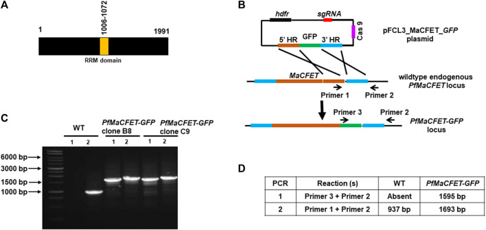
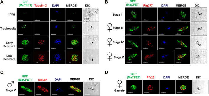
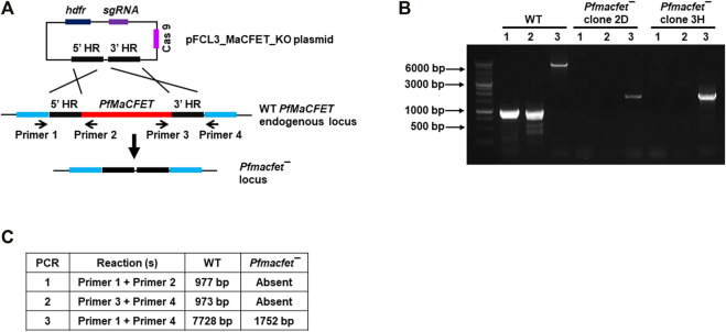
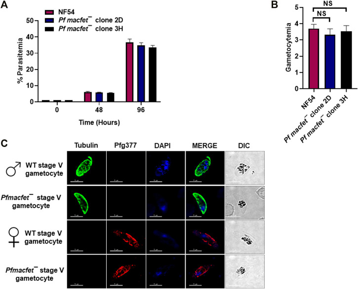
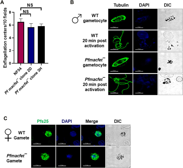
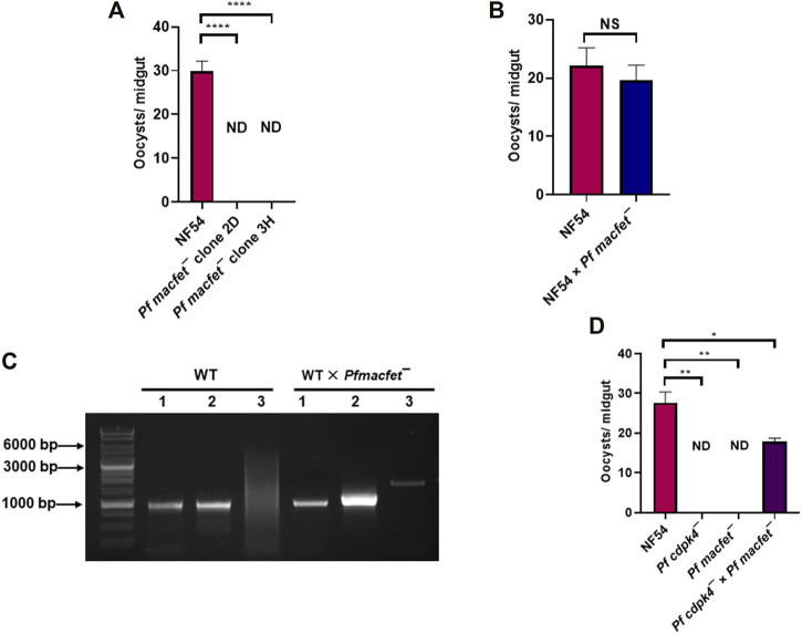
Plasmodium falciparum sexual stage gametocytes are critical for parasite transmission from the human host to the mosquito vector. Mature gametocytes generate fertile male (micro-) or female (macro-) gametes upon activation inside the mosquito midgut. While a number of parasite genes have been described that are critical for P. falciparum gametogenesis and fertility, no parasite gene has been shown to have a unique function in macrogametes. The genome of P. falciparum encodes numerous RNA-binding proteins. We identified a novel protein containing a putative RNA-binding domain, which we named Macrogamete-Contributed Factor Essential for Transmission (MaCFET). This protein is expressed in the asexual and sexual stages. Parasites that carry a deletion of MaCFET (Pfmacfet¯), developed normally as asexual stages, indicating that its function is not essential for the asexual proliferation of the parasite in vitro. Furthermore, Pfmacfet¯ male and female gametocytes developed normally and underwent activation to form microgametes and macrogametes. However, by utilizing genetic crosses, we demonstrate that Pfmacfet¯ parasites suffer a complete female-specific defect in successful fertilization. Therefore, PfMaCFET is a critical female-contributed factor for parasite transmission to the mosquito. Based on its putative RNA-binding properties, PfMaCFET might be in involved in the regulation of mRNAs that encode female-specific functions for fertilization or female-contributed factors needed post fertilization.

Keywords: fertility; gamete; gametocyte; mosquito; transmission.


Conflict of interest statement

The authors declare that the research was conducted in the absence of any commercial or financial relationships that could be construed as a potential conflict of interest.

Figures

FIGURE 1
FIGURE 1
Generation of PfMaCFET-GFP parasites. (A) Schematic for PfMaCFET domains showing an RNA recognition motif (RRM) domain. (B) Schematic for endogenous tagging of the PfMaCFET locus with GFP. The pFCL3_MaCFET_GFP plasmid contains homology arms from the 5′ (5′HR) and 3′ (3′HR) regions of the PfMaCFET locus, a single guide RNA seq (sgRNA), Cas9 and human dihydrofolate reductase (hDHFR). (C) Confirmation of PfMaCFET-GFP parasite generation by genotyping PCR. The oligonucleotides were designed and positions are indicated by arrows in (B) to confirm the introduction of the GFP tag. (D) The expected amplicon sizes for different sets of PCR primer combinations are indicated.
FIGURE 2
FIGURE 2
Expression and localization of PfMaCFET in asexual and sexual stage parasites (A) IFAs were performed using thin culture smears of asexual stages (ring, trophozoite, early and late schizonts), (B) sexual (stage II-V gametocytes) stages, (C) male gametocyte, (D) female gamete using anti-GFP antibody (in green) in combination with anti-Tubulin X (A), anti-PfG377 (B), anti-Tubulin (C) anti-Pfs25 (D). The parasite DNA was visualized with DAPI (blue). Representative images are shown. DIC, differential interference contrast. DAPI, 4′,6-diamidino-2-phenylindole, Scale bar = 5 μm.
FIGURE 3
FIGURE 3
Generation of Pfmacfet¯ parasites. (A) For generation of Pfmacfet¯ parasites a similar strategy as described in Figure 1 was used. pFCL3_PfMaCFET_KO plasmids had homology regions from 5′ (5′HR) and 3′ (3′HR) of the PfMaCFET locus, a single guide RNA seq (sgRNA) and human dihydrofolate reductase (hDHFR). (B,C) PfMaCFET deletion confirmation via diagnostic PCR. The oligonucleotides were designed from outside the 5′HR and 3′HR and the PfMaCFET locus and positions are indicated by arrows in (A). The expected amplicon sizes for different set of PCR primer combinations are indicated in (B,C).
FIGURE 4
FIGURE 4
Pfmacfet¯ parasites grow normally as asexual parasites and undergo gametocytogenesis. (A) Pfmacfet¯ (clone 2D and 3H) parasites grew at a similar rate as WT PfNF54. Data were averaged from three biological replicates and presented as the mean ± standard deviation (SD). (B) Bar graph shows gametocytemia for WT PfNF54 and Pfmacfet¯ parasites (clone 2D and 3H) measured on day 15. Data were averaged from three biological replicates and presented as the mean ± standard deviation (SD). (C) IFAs performed on thin blood culture smears for WT PfNF54 or Pfmacfet¯ (clone 3H) using α-tubulin (green), a male-specific marker, and Pfg377 (green), a marker for female gametocytes. Representative images are shown. DIC, differential interference contrast. DAPI, 4′,6-diamidino-2-phenylindole, Scale bar = 5 μm.
FIGURE 5
FIGURE 5
Pfmacfet¯ parasites form male and female gametes. (A) Number of exflagellation centers (vigorous flagellar beating of microgametes in clusters of RBCs) per microscopic field at 15 min post-activation. Data were averaged from three biological replicates and presented as the mean ± standard deviation (SD). (B) IFAs performed on thin blood culture smears for gametocytes activated for 20 min in vitro for WT PfNF54 or Pfmacfet¯ (clone 3H) using α-tubulin II (green), a male-specific marker. α-Tubulin II staining showed microgametes emerging from exflagellating male gametocyte in both WT PfNF54 and Pfmacfet¯ gametocytes. (C) IFAs performed on thin blood culture smears for day 15 stage V gametocytes for WT PfNF54 or Pfmacfet¯ (clone 3H) using α-Pfs25 (green), a marker for female gametes. Representative images are shown. DIC, differential interference contrast. DAPI, 4′,6-diamidino-2-phenylindole, Scale bar = 5 μm.
FIGURE 6
FIGURE 6
PfMaCFET regulates fertility of female gametes. (A) Mosquitoes with single parasite lines or genetic crosses were dissected on day 7 post feed and number of oocysts were measured per midgut. Data were averaged from three biological replicates with a minimum of 50 mosquito guts and presented as the mean ± standard deviation (SD) n = 3. ****p-value= <0.0001. (B) Oocyst numbers for WT PfNF54 and the WT Pf NF54 × Pfmacfet¯ cross. Error bar indicates mean ± SD; n = 2. NS-Not significant. (C) Confirmation of transmission of Pfmacfet¯ parasites in a WT × Pfmacfet¯ clone 3H genetic cross by genotyping PCR. The oligonucleotides were designed from outside the 5′ and 3′ homology regions of the PfMaCFET locus and positions as indicated by arrows in Figure 3A. (D) Oocyst formation for WT PfNF54, Pfmacfet¯, Pfcdpk4¯ single parasite infections and the Pfmacfet¯ × Pfcdpk4¯ genetic cross infection. Error bar indicates mean ± SD; n = 2. ND-Not detected, **p-value = 0.0048, *p-value = 0.0399.

References

    1. Andreadaki M., Hanssen E., Deligianni E., Claudet C., Wengelnik K., Mollard V., et al. (2018). Sequential Membrane Rupture and Vesiculation during Plasmodium Berghei Gametocyte Egress from the Red Blood Cell. Sci. Rep. 8, 3543. 10.1038/s41598-018-21801-3 - DOI - PMC - PubMed
    1. Billker O., Dechamps S., Tewari R., Wenig G., Franke-Fayard B., Brinkmann V. (2004). Calcium and a Calcium-dependent Protein Kinase Regulate Gamete Formation and Mosquito Transmission in a Malaria Parasite. Cell 117, 503–514. 10.1016/s0092-8674(04)00449-0 - DOI - PubMed
    1. Billker O., Lindo V., Panico M., Etienne A. E., Paxton T., Dell A., et al. (1998). Identification of Xanthurenic Acid as the Putative Inducer of Malaria Development in the Mosquito. Nature 392, 289–292. 10.1038/32667 - DOI - PubMed
    1. Boag P. R., Atalay A., Robida S., Reinke V., Blackwell T. K. (2008). Protection of Specific Maternal Messenger RNAs by the P Body Protein CGH-1 (Dhh1/RCK) during Caenorhabditis elegans Oogenesis. J. Cel. Biol. 182, 543–557. 10.1083/jcb.200801183 - DOI - PMC - PubMed
    1. Bunnik E. M., Batugedara G., Saraf A., Prudhomme J., Florens L., Le Roch K. G. (2016). The mRNA-Bound Proteome of the Human Malaria Parasite Plasmodium Falciparum. Genome Biol. 17, 147. 10.1186/s13059-016-1014-0 - DOI - PMC - PubMed

LinkOut - more resources