. 2022 Mar 22;25(4):104150.
doi: 10.1016/j.isci.2022.104150. eCollection 2022 Apr 15.

Systematic characterization of wing mechanosensors that monitor airflow and wing deformations

Affiliations

Systematic characterization of wing mechanosensors that monitor airflow and wing deformations

Joseph Fabian et al. iScience. .

Abstract

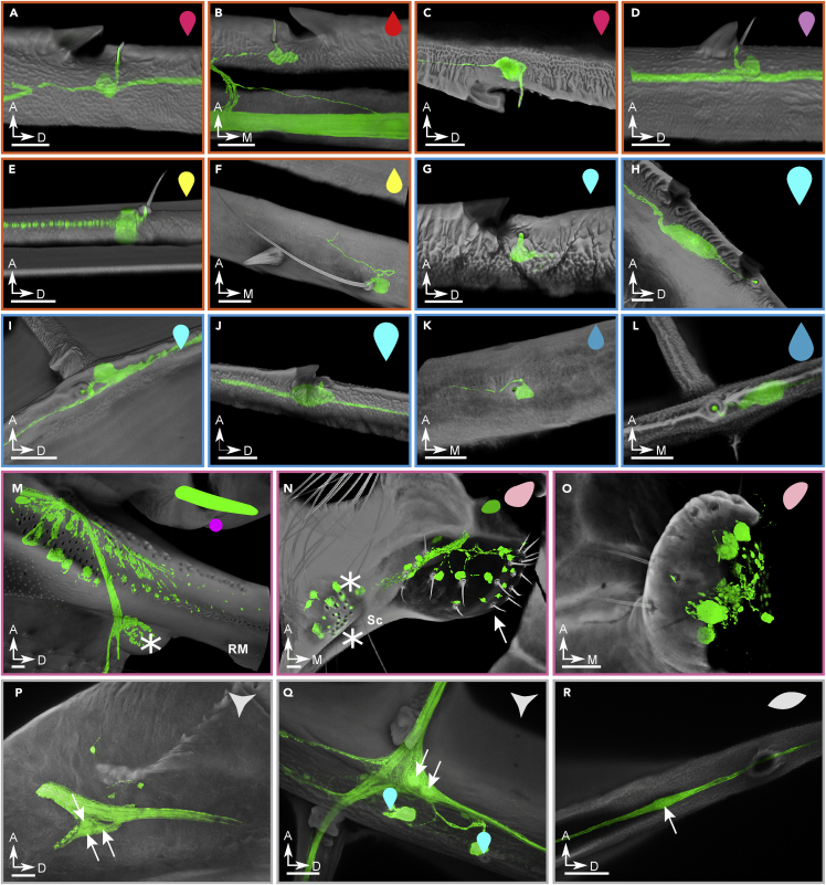
Animal wings deform during flight in ways that can enhance lift, facilitate flight control, and mitigate damage. Monitoring the structural and aerodynamic state of the wing is challenging because deformations are passive, and the flow fields are unsteady; it requires distributed mechanosensors that respond to local airflow and strain on the wing. Without a complete map of the sensor arrays, it is impossible to model control strategies underpinned by them. Here, we present the first systematic characterization of mechanosensors on the dragonfly's wings: morphology, distribution, and wiring. By combining a cross-species survey of sensor distribution with quantitative neuroanatomy and a high-fidelity finite element analysis, we show that the mechanosensors are well placed to perceive features of the wing dynamics relevant to flight. This work describes the wing sensory apparatus in its entirety and advances our understanding of the sensorimotor loop that facilitates exquisite flight control in animals with highly deformable wings.

Keywords: Animal physiology; Biomechanics; Entomology; Sensory neuroscience.


Conflict of interest statement

The authors declare no competing interests.

Figures

None
Graphical abstract
Figure 1
Figure 1
The axon routing pattern of selected odonates Neuronal innervation diagram of male eastern amberwing dragonfly (Perithemis tenera) forewing (A), hindwing (B), and a male blue-fronted dancer damselfly (Argia apicalis) hindwing (C). A dedicated afference wing nerve branches out into different veins from the wing base. The color tones of axonal tracks were chosen arbitrarily for readability. They do not imply hierarchy or relationship of the tracks. Dots indicate terminal mechanosensory cell body for each track; dashed lines denote merging tracks; dotted lines represent tracks that were interpolated from incomplete back-fill images and mechanosensors found on the veins. Major veins and structural elements of the wings are labeled: C- costa; Sc - subcosta; RM - radius/media; Cu - cubitus; (A) anal; Arc - arculus; T - triangle; ST - supratriangle; Q - quadrilateral. The corrugations of the wings/topology of veins are represented by “+” for “ridges” (dorsally convex) and “-” for “valleys” (dorsally concave). See also Figure S1.
Figure 2
Figure 2
Variations in the axon routing patterns of dragonfly wings (A) Variability of axonal tracks in two dragonflies (Perithemis tenera) fore- and hindwings. The dragonfly II wings were superimposed on the dragonfly I’s, scaled, and warped in Adobe illustrator to align the wing margins and main veins. Each vein carries a single nerve. (B) Variability of the axonal track passage at the nodus of P. tenera: axons of the leading-edge wing margin sensors distal to nodus may continue down the costa or take an alternative path down the radius vein. The path does not seem to depend on the sex of the insect and the variability occurs in both fore (F) and hind (H) wings. (C) Real paths (red) and minimal distance paths (green) of axons innervating a wing margin bristle-bump complex neuron (anterior pair) and a campaniform sensillum (posterior pair) in a male (dragonfly I) forewing. (D) The real path distance and minimal path distance for 31 pseudorandomly selected sensors on the forewing and hindwing.
Figure 3
Figure 3
The classification and morphology of all wing sensors Examples of sensors found on the wings of the dragonfly Perithemis tenera. The scale bars are all 25 μm. (A-F) Candidate airflow sensors: (A) (B) (C) costa dorsal, costa ventral, and trailing-edge margin bristle complex. These three special examples show the double-innervated bristle which consists <25% of all the wing margin bristles. All other bristles have one dendrite innervating the bristle base (not shown). (D) Radius bristle-bump; all bristles of this type are innervated by one neuron at the base. (E) Short bristle of the type present on several major veins. (F) Long bristle of the type present on the medial part of major veins. (G–L) Strain sensors: (G) dorsal costa campaniform sensillum (CS); (H) large dorsal costa CS; (I) dorsal cross vein CS; (J) large radius anterior CS immediately distal to pterostigma; (K) ventral subcosta CS; (L) terminal ventral CS of the radius posterior 1 (RP1) vein. (M–O) Wing base sensory fields: (M) crevice organ; two parallel fields of directionally tuned elliptical CS at the base of radius/media (RM) vein. Asterisk marks the dorsal insertion site of a wing base chordotonal organ; (N) hair plate (arrow) and two adjacent CS fields (asterisks) ventrally at the base of subcosta; (O) hair plate ventrally at the base of cubitus. (P–R) Multipolar and bipolar receptors: (P) multipolar receptor at the base of costa; arrows point to/indicate cell bodies; (Q) multipolar receptor located at the junction of the anal vein and the first/medial cross vein connecting it to cubitus. Two adjacent dorsal CS are indicated with cyan markers. (R) Bipolar receptor. All example images are from the right forewing, with cuticle in gray, and neurons in green. The symbols in the upper right corner of each panel are notations to show sensor type and dorsal/ventral placement which will be used in Figuers 4 and 5. See also Figure S2.
Figure 4
Figure 4
A sensory map of odonate wings Distribution of all confirmed sensors on the Perithemis tenera dragonfly fore- (A) and hind (B) wings and a hindwing of Argia apicalis damselfly (C). All sensor notations follow Figure 3 and the figure legend. Inset (i): maximum intensity projection showing P. tenera right forewing base dorsally and ventrally with the sensor fields outlined. Insets (ii) and (iii): diagrams showing the wings’ natural resting sweep angles; the red reference lines mark the wing span axis perpendicular to the anatomical wing hinges.
Figure 5
Figure 5
Quantifying the cell body volume and axon diameter of wing neurons (A) Positions of wing sensors (selected to evenly sample the wingspan) on the right hindwing of Perithemis tenera. Cyan—campaniform sensilla (CS) orange—bristle-bump complexes. (B) Example images of the cell body and axon of wing sensors, with cuticle in green and neurons in red. Grayscale images underneath show the neuron channel alone. (C) Cell body volume for wing sensors outlined in (A). (D and E) Axon diameter for bristle-bumps (D) and campaniform sensilla (E), black line indicates the axon diameter required to achieve equal response latency for different axon lengths, dashed line indicates simple linear fit.
Figure 6
Figure 6
Positions of campaniform sensilla at the wing tips of studied odonates Campaniforms on costa (red), radius anterior (dark orange), and radius posterior (light orange) are marked. Dots denote dorsal, crosses – ventral sensors. Enlarged markers indicate campaniform sensilla innervated by sensory neurons possessing significantly larger soma as seen in backfills for Argia apicalis and Perithemis tenera, and inferred from a prominent bulge on the vein in Ischnura verticalis, likely expanded to accommodate a large cell body. Scale bars: 1 mm.
Figure 7
Figure 7
A comparison of selected sensor distribution across odonate families Sensor density distribution (A–H) and sensor count (I–K) across 15 odonate species. Normalized sensor density is shown for dorsal and ventral side over normalized sensor position on three longitudinal veins, costa (A and B), subcosta (C–E), and radius (F–H) (See Table S1 for sensor count values). The insets in each panel highlight sensor type and the respective vein. Sensor density is normalized by the mean of each density curve. Sensor position is normalized by the length of the respective right forewing. The black bold line shows the mean of all species in each plot. (I–K) Sensor count is the sum for each sensor type of all three longitudinal veins. The sensor count is plotted over the respective wing length for each species. Black line shows the ordinary least squares (OLS) regression and the gray shaded area shows the 95% confidence interval. Each data point is color coded by the respective species (see legend in upper right panel).
Figure 8
Figure 8
Strain field propogation on a simulated flapping wing (A–D) The hindwing of a Sympetrum striolatum was reconstructed in 3D for this structural analysis. Four time instances of the flapping cycle are presented: beginning of downstroke (A), mid-downstroke (B), beginning of upstroke (C), and mid-upstroke (D). A hindwing of a Sympetrum striolatum was reconstructed in 3D for this simulation. (E and F) Spanwise strain along ventral subcosta and dorsal radius where 962 campaniform sensilla can be found. The color map represents different phase of a flapping cycle. (G and H) Spanwise strains along the veins, each line is normalized by the mean of the strain along the vein at that time. The x axis (path length, or position along vein) for the spanwise strains are normalized with the total path length for the corresponding vein. (I and J) Temporal variation of the mean spanwise strain on the ventral subcosta and dorsal radius. The measured positions of campaniform sensilla are shown by magenta dots (E–H). The vertical variation of the dots is purely for readability. See also Figure S3.

Similar articles

Cited by

References

    1. Ai H., Yoshida A., Yokohari F. Vibration receptive sensilla on the wing margins of the silkworm moth Bombyx mori. J. Insect Physiol. 2010;56:236–246. doi: 10.1016/j.jinsphys.2009.10.007. - DOI - PubMed
    1. Albert P.J., Zacharuk R.Y., Wong L. Structure, innervation, and distribution of sensilla on the wings of a grasshopper. Can. J. Zool. 1976;54 doi: 10.1139/z76-178. 1542–53. - DOI
    1. Bomphrey R.J., Godoy-Diana R. Insect and insect-inspired aerodynamics: unsteadiness, structural mechanics and flight control. Curr. Opin. Insect Sci. 2018;30:26–32. doi: 10.1016/j.cois.2018.08.003. - DOI - PMC - PubMed
    1. Bomphrey R.J., Nakata T., Henningsson P., Lin H.-T. Flight of the dragonflies and damselflies. Philos. Trans. R. Soc. Lond. B Biol. Sci. 2016;371 doi: 10.1098/rstb.2015.0389. - DOI - PMC - PubMed
    1. Boyde A., Mccorkell F.A., Taylor G.K., Bomphrey R.J., Doube M. Iodine vapor staining for atomic number contrast in backscattered electron and X-ray imaging. Microsc. Res. Tech. 2014;77:1044–1051. doi: 10.1002/jemt.22435. - DOI - PMC - PubMed

LinkOut - more resources