. 2022 Apr 8:16:875726.
doi: 10.3389/fncel.2022.875726. eCollection 2022.

The T-Type Calcium Channel Cav3.2 in Somatostatin Interneurons in Spinal Dorsal Horn Participates in Mechanosensation and Mechanical Allodynia in Mice

Affiliations

The T-Type Calcium Channel Cav3.2 in Somatostatin Interneurons in Spinal Dorsal Horn Participates in Mechanosensation and Mechanical Allodynia in Mice

Yu-Ru Zhi et al. Front Cell Neurosci. .

Abstract

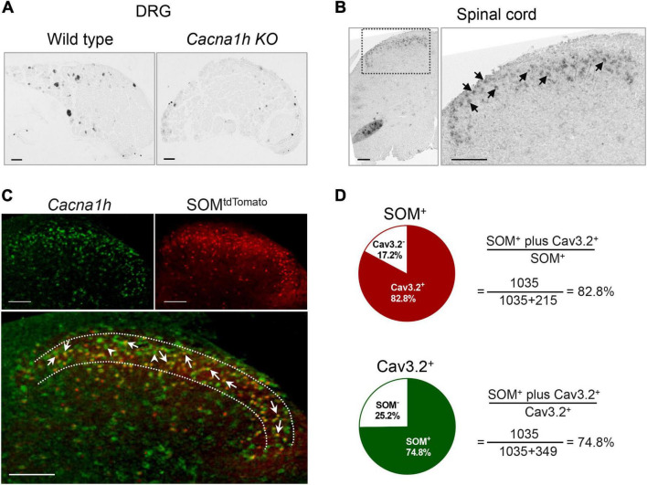
Somatostatin-positive (SOM+) neurons have been proposed as one of the key populations of excitatory interneurons in the spinal dorsal horn involved in mechanical pain. However, the molecular mechanism for their role in pain modulation remains unknown. Here, we showed that the T-type calcium channel Cav3.2 was highly expressed in spinal SOM+ interneurons. Colocalization of Cacna1h (which codes for Cav3.2) and SOM tdTomato was observed in the in situ hybridization studies. Fluorescence-activated cell sorting of SOM tdTomato cells in spinal dorsal horn also proved a high expression of Cacna1h in SOM+ neurons. Behaviorally, virus-mediated knockdown of Cacna1h in spinal SOM+ neurons reduced the sensitivity to light touch and responsiveness to noxious mechanical stimuli in naïve mice. Furthermore, knockdown of Cacna1h in spinal SOM+ neurons attenuated thermal hyperalgesia and dynamic allodynia in the complete Freund's adjuvant-induced inflammatory pain model, and reduced both dynamic and static allodynia in a neuropathic pain model of spared nerve injury. Mechanistically, a decrease in the percentage of neurons with Aβ-eEPSCs and Aβ-eAPs in superficial dorsal horn was observed after Cacna1h knockdown in spinal SOM+ neurons. Altogether, our results proved a crucial role of Cav3.2 in spinal SOM+ neurons in mechanosensation under basal conditions and in mechanical allodynia under pathological pain conditions. This work reveals a molecular basis for SOM+ neurons in transmitting mechanical pain and shows a functional role of Cav3.2 in tactile and pain processing at the level of spinal cord in addition to its well-established peripheral role.

Keywords: SOM neurons; intraspinal injection; knockdown; low-voltage activated calcium channel; spinal cord slice recording.


Conflict of interest statement

The authors declare that the research was conducted in the absence of any commercial or financial relationships that could be construed as a potential conflict of interest.

Figures

FIGURE 1
FIGURE 1
In situ hybridization staining of Cacna1h in DRG and spinal dorsal horn. (A) In situ hybridization staining of Cacna1h in the DRG of wild type (WT, left) and Cacna1h knockout (KO, right) mice. (B) In situ hybridization staining of Cacna1h in the lumbar segment of the spinal cord in mice. Arrows indicate Cacna1h+ cells. (C) In situ hybridization staining of Cacna1h (left) and SOMtdTomato neurons (right) in the spinal dorsal horn. Superimposition of the images is shown below. Arrows indicate Cacna1h and SOMtdTomato double-positive cells, and arrowheads indicate Cacna1h-positive cells. (D) Quantification analysis of the proportion of double-positive cells in SOM+ (top) and Cav3.2+ (below) cells. Sixteen hemisections of the spinal cord from 4 male mice in each group were quantified. Scale bar, 100 μm.
FIGURE 2
FIGURE 2
High expression of Cacna1h in SOM+ neurons in spinal dorsal horn. (A) Representative image of SOMtdTomato neurons in sagittal sections of the spinal cord. Right image is an enlarged view of the boxed area. Scale bar (left), 500 μm. Scale bar (right), 50 μm. (B) Sorting of SOMtdTomato neurons in the spinal dorsal horn by flow cytometry. Spinal dorsal horn neurons from SOMCre mice were used as controls (left). The P4 region represents Tomato-positive neurons, whereas the P5 region represents Tomato-negative neurons. (C,D) qPCR analysis of Sst (C) and Cacna1h (D) gene expression levels in Tomato-negative and Tomato-positive cells. *P < 0.05, **P < 0.01, Student’s unpaired t-test. FSC, forward scatter. SSC, side scatter.
FIGURE 3
FIGURE 3
Knockdown of Cacna1h in spinal SOM+ neurons does not affect motor ability or thermal sensations in naïve mice. (A) Schematic illustration of viral-mediated knockdown of Cacna1h in spinal SOM+ neurons through intraspinal injection. Non-silence shRNA virus was used as a control. (B) Expression of the GFP-tagged virus in the spinal cord. Scale bar, 100 μm. (C) qPCR analysis of the relative expression level of Cacna1h 28 d after injection of Cacna1h knockdown virus. Non-silence shRNA virus was used as a control. (D–H) Behavioral tests of motor ability and thermal sensations in the mice after knockdown of Cacna1h in spinal SOM+ neurons. The total distance traveled in the open field (D), the latency to fall in the rotarod (E), the behavioral responses to acetone-evoked cooling (F), the withdrawal latency in the Hargreaves test (G) and the licking latency in the hot plate at 54°C (H) were unaffected by Cacna1h knockdown in spinal SOM+ neurons. Student’s unpaired t-test.
FIGURE 4
FIGURE 4
Knockdown of Cacna1h in spinal SOM+ neurons decreases the response to light touch and noxious mechanical stimulation in naïve mice. (A–C) Behavioral tests of the tactile sensation. Decreased the response to brush stimulation (A) and “pulled out” cotton swabs (B), and increased licking latency to sticky tape (C) were observed after Cacna1h knockdown in spinal SOM+ neurons. (D) The 50% withdrawal threshold to von Frey filament stimulation was unaffected by Cacna1h knockdown in spinal SOM+ neurons. (E) The percentage of response to pinprick stimulation was reduced after Cacna1h knockdown in spinal SOM+ neurons. *P < 0.05, Student’s unpaired t-test. (F) The licking latency to pinch stimulation was not affected by Cacna1h knockdown in spinal SOM+ neurons. *P < 0.05, **P < 0.01, Student’s unpaired t-test.
FIGURE 5
FIGURE 5
Knockdown of Cacna1h in spinal SOM+ neurons attenuated heat hyperalgesia and dynamic allodynia but not static allodynia in a CFA-induced chronic inflammatory pain model in mice. (A) Schematic illustration of the timeline of virus injection, model establishment and behavioral tests. Non-silence shRNA virus was used as a control. (B) The withdrawal latency in the Hargreaves test was increased after Cacna1h knockdown in spinal SOM+ neurons in the CFA model. ### P < 0.001, two-way ANOVA with Sidak post hoc analysis. **P < 0.01, difference between the two groups at the corresponding time points. (C) Licking latency in the hot plate of 54°C was elongated by Cacna1h knockdown in spinal SOM+ neurons at 7 days post CFA injection. *P < 0.05, Student’s unpaired t-test. (D,E) The dynamic allodynia score was reduced (D), but the static allodynia reflected as the 50% withdrawal threshold was not affected (E) by Cacna1h knockdown in spinal SOM+ neurons in the CFA model. ### P < 0.001, two-way ANOVA with Sidak post hoc analysis. *P < 0.05, **P < 0.01, difference between the two groups at the corresponding time points. BL, baseline.
FIGURE 6
FIGURE 6
Knockdown of Cacna1h in spinal SOM+ neurons attenuated dynamic and static allodynia in an SNI-induced neuropathic pain model in mice. (A) Schematic illustration of the timeline of virus injection, model establishment and behavioral tests. Non-silence shRNA virus was used as a control. (B,C) Both the dynamic allodynia score (B) and the static allodynia reflected as the 50% withdrawal threshold (C) were reduced by Cacna1h knockdown in spinal SOM+ neurons in the SNI model. ### P < 0.001, two-way ANOVA with Sidak post hoc analysis. *P < 0.05, **P < 0.01, ***P < 0.001, difference between the two groups at the corresponding time points. BL, baseline.
FIGURE 7
FIGURE 7
Knockdown of Cacna1h in spinal SOM+ neurons partially closed the superficial Aβ pathway in neuropathic pain. (A) Schematic drawing of patch clamp recordings on parasagittal spinal cord slice with dorsal root and DRG attached and recorded neurons (red dots). (B) Representative traces and percentage of neurons in laminae I-IIo with Aβ-eEPSC (left), Aβ-eAP (middle) and Aβ-eIPSC (right) in naïve mice under normal ACSF, ACSF containing bicuculline (Bic) plus strychnine (Stry), non-silence-shRNA-injected SOMCre mice with SNI (shControl&SNI) and Cacna1h-shRNA-injected SOMCre mice with SNI (shCacna1h&SNI). All data are represented as percentage. *P < 0.05. **P < 0.01. ***P < 0.001. Chi-square test.
FIGURE 8
FIGURE 8
Cav3.2 channels in spinal SOM+ neurons are involved in the sensory processing of light touch in the basal state and contribute to heat hyperalgesia and mechanical allodynia in pathological pain, including inflammatory pain and neuropathic pain. In the current work, we demonstrated that the Cacna1h gene was enriched in SOM+ neurons in the spinal dorsal horn. After knockdown of Cacna1h expression in spinal SOM+ neurons by intraspinal injection of Cre-dependent virus, the mice displayed a lower response to light touch, including brush, cotton and tape stimulation, and showed a decreased response to the noxious mechanical stimulation in the basal state. In the pathological state, knockdown of Cacna1 h in spinal SOM+ neurons attenuated thermal hyperalgesia and dynamic mechanical allodynia behaviors in the inflammatory pain model and both dynamic and static mechanical allodynia behaviors in the neuropathic pain model.

Similar articles

Cited by

References

    1. Bernal Sierra Y. A., Haseleu J., Kozlenkov A., Begay V., Lewin G. R. (2017). Genetic tracing of Cav3.2 T-type calcium channel expression in the peripheral nervous system. Front. Mol. Neurosci. 10:70. 10.3389/fnmol.2017.00070 - DOI - PMC - PubMed
    1. Bouhassira D., Attal N., Alchaar H., Boureau F., Brochet B., Bruxelle J., et al. (2005). Comparison of pain syndromes associated with nervous or somatic lesions and development of a new neuropathic pain diagnostic questionnaire (DN4). Pain 114 29–36. 10.1016/j.pain.2004.12.010 - DOI - PubMed
    1. Bourinet E., Alloui A., Monteil A., Barrere C., Couette B., Poirot O., et al. (2005). Silencing of the Cav3.2 T-type calcium channel gene in sensory neurons demonstrates its major role in nociception. EMBO J. 24 315–324. 10.1038/sj.emboj.7600515 - DOI - PMC - PubMed
    1. Boyle K. A., Gradwell M. A., Yasaka T., Dickie A. C., Polgár E., Ganley R. P., et al. (2019). Defining a spinal microcircuit that gates myelinated afferent input: implications for tactile allodynia. Cell Rep. 28 526–540. 10.1016/j.celrep.2019.06.040 - DOI - PMC - PubMed
    1. Cain S. M., Snutch T. P. (2010). Contributions of T-type calcium channel isoforms to neuronal firing. Channels 4 475–482. 10.4161/chan.4.6.14106 - DOI - PMC - PubMed

LinkOut - more resources